
Comparison of two techniques for measuring pulse wave velocity and central blood pressure
- DOI
- 10.1016/j.artres.2011.06.001How to use a DOI?
- Keywords
- Pulse wave velocity; Augmentation index; Methods; SphygmoCor; Arteriograph
- Abstract
Pulse wave velocity (PWV) is an important parameter in the assessment of overall cardiovascular risk and there is well documented predictions of mortality in special groups of patients. Several methods are available for measuring the velocity and other parameters of the pulse wave. Our aim was to assess the comparability of two methods; applanation tonometry, which is used by the SphygmoCor device and oscillometry employed by the Arteriograph system. Published data on their comparability are contradictory. Thirty-three patients of both sexes were examined in the study. Mean PWV was significantly higher with Arteriograph than SphygmoCor (10.2 m/s ± 3.9 vs. 8.9 m/s ± 2.5) and Aix was significantly higher with Arteriograph than SphygmoCor too (29.3 ± 16.3 vs. 21.2 ± 12.6). The lack of agreement between the two methods is confirmed also by the Bland–Altman plot. Due to working principle of the Arteriograph possible conclusion is that parameters provided by the Arteriograph are the measures of brachial stiffness and not aortic stiffness. However, the method used by the Arteriograph is definitely much simpler and more time-efficient than applanation tonometry used by the SphygmoCor device.
- Copyright
- © 2011 Association for Research into Arterial Structure and Physiology. Published by Elsevier B.V. All rights reserved.
- Open Access
- This is an open access article distributed under the CC BY-NC license.
Introduction
Pulse wave velocity (PWV) is an indicator of structural changes of the aorta and large vessels. In the current guidelines for the management of patients with arterial hypertension,1–3 PWV is an important parameter in the assessment of overall cardiovascular risk and there is well documented predictions of mortality in special groups of patients, eg. elderly in patients with end-stage renal disease, diabetes mellitusor hypertension and in the general population as well4 and is correlated with coronary heart disease.5 Measurement of carotid-femoral pulse vawe velocity is the only non-invasive method which is acuurate enough to be used as a diagnostic procedures in a clinical setting. High values indicate stiffness of arteries.1
Augmentation and the augmentation index (AIx) are being used as parameters of wave reflection. They are a direct measure of wave reflection. The effect of wave reflection on the aortic systolic pressure peak can be described as augmentation. Dividing the augmentation pressure by the pulse pressure gives the AIx. Ithas also been shown that AIx is closely correlated with thecardiovascular risk and correlates with left ventricular mass even in normotensive subjects.6 Both PWV and augmentation provide extensive informationon the arterial vascular system.
Several methods are available for measuring the velocity and other parameters of the pulse wave (central blood pressure, AIx). The oldest is applanation tonometry, which is used by the SphygmoCor device (At Cor Medical, Australia). Pulse wave velocity is calculated as the ratio of the distance between carotid artery and femoral artery, and the pulse transit time from one location to the other. Another method is oscillometry employed by the Arteriograph system (TensioMed, Hungary). This device measures the pulse wave at one single location using brachial cuff that is overinflated to 35 mmHg over systolic blood pressure. The time difference between the first systolic wave and the second reflected wave is used as a transit time. Pulse wave velocity can be calculated as the ration of travelled distance between jugulum and symphysis over transit time.
Our aim was to assess the comparability of these two methods since the published data on their comparability are contradictory.
Methods
All measurements were performed by the same technician in the same room at a constant temperature and were unaffected by external environmental influences.
Blood pressure (BP) was measured at the upper arm using an OMRON 705IT oscillometric monitor in accordance with the international and national guidelines.2,3 The first measurement in each patient was performed by applanation tonometry, as a standard method, using the SphygmoCor device. Pulse curves were recorded over the radial, carotid and femoral arteries, and the ECG was recorded simultaneously. The pulse pressure curve measured at the radial artery was calibrated against an oscillometrically measured upper-arm blood pressure. The augmentation index (AIx) was then determined by calculating the aortic pressures by means of a transfer function.7 PWV was calculated from the pulse waves curves recorded over the carotid and femoral arteries sequentially and the simultaneously obtained ECG tracings. The distance between the carotid registration site and the femoral arterial registration site was then determined, and the delay of the pulse wave relative to the QRS complex in the ECG are the basis for PWV calculation.8
The measurement with the Arteriograph was performed directly after the initial measurement. In this method, a cuff of appropriate size is wrapped around the upper arm and overinflated to 35 mmHg over the systolic blood pressure. Oscillations in the cuff are a reflexion of oscillations of the brachial artery. The principle of the oscillometric method is based on plethysmography and registers pulsatile pressure changes in an artery. The software of the Arteriograph decomposes the early, late systolic and diastolic waves and also determines the onset and the peaks of the waves and than calculates PWV, assuming that the measuured external distance from jugulum to symphysis is an approximation of the distance to the reflection site. The augmentation index corresponds to the pressure difference between the first and second wave in relation to the pulse pressure.9
The study was conducted on random patients scheduled for PWV evaluation. Thirty-three patients of both sexes were examined in the study. As shown in Table 1, the subjects differed considerably in age, body mass index (BMI) and BP.
| N | Minimum | Maximum | Mean | Std. deviation | |
|---|---|---|---|---|---|
| Age | 33 | 24.00 | 85.00 | 54.4242 | 15.43946 |
| BMI | 33 | 19.94 | 42.53 | 28.8941 | 4.35818 |
| SBP (mmHg) | 33 | 97 | 192 | 143.79 | 20.320 |
| DBP (mmHg) | 33 | 60 | 116 | 81.79 | 13.233 |
| PP (mmHg) | 33 | 35.00 | 101.00 | 62.0000 | 15.04992 |
BMI: body mass index; SBP: systolic blood pressure; DBP: diastolic blood pressure; PP: pulse pressure.
Basic characteristics of the study subjects.
All values are given as a mean and standard deviation. Differences between two groups were testedwith the t-test. Less than 5% risk was taken to besignificant. The variance was plotted according to Bland and Altman.10
Results
Comparison of the results of the PWV measurements (made with the Arteriograph and the SphygmoCor device) is presented in Table 2. The difference between the two methods is statistically significant both for PWV and for the calculated AIx values. The lack of agreement between the two methods is confirmed also by the Bland–Altman plot, both for PWV and AIx (Figs. 1 and 2). The average discrepancy between the results obtained with the two methods is greater for AIx than for PWV.
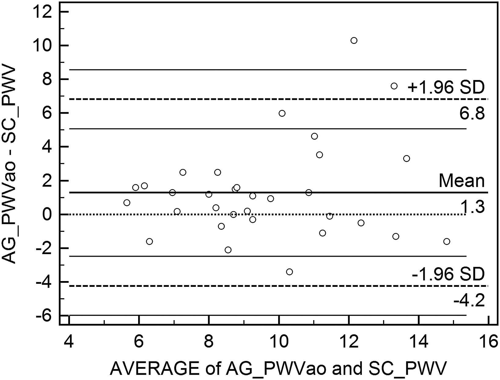
Bland–Altman plot of differences between the results for pulse wave velocity obtained with two methods.

Bland–Altman plot of differences between augmentation index values obtained with two methods.
| Mean | N | SD | Sig. | |
|---|---|---|---|---|
| SphygmoCor PWV | 8.916 | 32 | 2.5391 | 0.004 |
| Arteriograph PWV | 10.2075 | 32 | 3.03080 | |
| SphygmoCor AIx | 21.13 | 32 | 12.569 | 0.000 |
| Arteriograph AIx | 29.2800 | 32 | 16.31157 |
PWV: pulse wave velocity; AIx: augmentation index.
Comparison of results for pulse wave velocity.
Discussion
Pulse wave velocity is a clinically accepted index for evaluation of aortic stiffness as a factor of cardiovascular risk.11 There is increasing evidence that central blood pressure is an important clinical parameter. It is affected by various drug regimens used in the treatment of hypertension and is not always equal to brachial BP.12,13 The recent publication of reference values for PWV and central BP14 has increased the clinical applicability of these measurements.
Applanation tonometry is currently the most widely used method for non-invasive evaluation of the shape of the pulse wave curve and calculation of parameters relevant for estimation of central BP. The method is technically very demanding. The subject must rest before the measurement, and the examiner must position the probe correctly over the artery and use the right amount of pressure to obtain adequate pulse wave recordings. Applying excessive pressure over the carotid artery may be fatal if an unstable atherosclerotic plaque happens to be present at that site. The oscillometric method used by the Arteriograph is much simpler to perform and requires less experience on the part of the examiner. Correct placement of the cuff on the upper arm is essential for accurate measurements. The examination is brief and provides simultaneous measurement of brachial BP, which must be determined separately when applanation tonometry is used.
Published data on the comparability of these two methods for the measurement of PWV and BP in the aorta differ considerably among studies. Some authors observed satisfactory agreement between the results15 and others did not.16 The advantages and shortcomings of each method are described in the literature.17 Our data suggest that the two methods are not interchangeable. It seems that the aortic PWV measured and calculated by Arteriograph represents PWV in the arm rather than aorta. Inflation of the brachial cuff to a suprasystolic pressure is the basic working principle of the Arteriograph which leads to an amplification of the second peak in the brachial pressure curve which is hardly noticable in normal conditions.18 We agree with the explanation that measured wave is predominantly the result of waves traveling back and forth in the braciahl artery reflected distally on the occluded cuff and proximally on the open-end reflection of the aortic junction.19 In some earlier studies it was shown that brachial PWV is higher than aortic, depending on blood pressure levels20,21 and possible peripheral vascular diseases of large arteries and not small arteries.22 However, the Arteriograph is certainly much simpler to use compared to the SphgymoCor system since the Arteriograph only measures at one location (the upper arm). Measurement can be done significantly faster.
Conclusion
Our results suggest, that the two methods cannot be used interchangeably either for measuring PWV or for determining AIx. PWV measured with Arteriograph is higher than PVW measured by SphygoCor system. Working principle of the Arteriograph leads us to the possible conclusion that parameter provided by the Arteriograph is a measure of brachial stiffness and not aortic stiffness.The method used by the Arteriograph is definitely much simpler and more time-efficient than applanation tonometry used by the SphygmoCor device. Probably, however, adjustment of the algorithms could bring the oscillometric method closer to applanation tonometry, which remains for the present the reference method.
References
Cite this article
TY  - JOUR
AU  - Rok Accetto
AU  - Barbara Salobir
AU  - Jana Brguljan
AU  - Primoz Dolenc
PY  - 2011
DA  - 2011/06/16
TI  - Comparison of two techniques for measuring pulse wave velocity and central blood pressure
JO  - Artery Research
SP  - 97
EP  - 100
VL  - 5
IS  - 3
SN  - 1876-4401
UR  - https://doi.org/10.1016/j.artres.2011.06.001
DO  - 10.1016/j.artres.2011.06.001
ID  - Accetto2011
ER  -