
P6.02 IN-VIVO ASSESSMENT OF THE ACCURACY OF CAROTID STRAIN ESTIMATES DERIVED FROM ULTRASONIC WALL TRACKING
- DOI
- 10.1016/j.artres.2011.10.088How to use a DOI?
- Open Access
- This is an open access article distributed under the CC BY-NC license.
Background: Ultrasonic wall tracking is a common tool in cardiovascular screening, where it is applied to assess arterial diameter distension waveforms. However, using multiple tracking points within the arterial wall and under the assumption of planar deformation, circumferential and radial strain can be obtained as εθθ=D/D and εrr=∂D/∂D.
Methods: We investigated the accuracy of these arterial strain estimates, using 10 representative data sets of the Asklepios population study . Tracking was done using a scanline perpendicular to the common carotid artery, far enough from the bulbus and showing clear intima-media and media-adventitia transitions. We started tracking from a designated point on the media-adventitia transition and from there on every 100 micrometer towards the lumen.
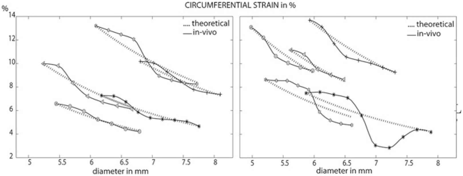
Results & Discussion: Data revealed an S-shaped curve of ϵθθ throughout the wall for all 10 subjects (fig.1, solid line), which was different from D/D calculated from conservation of mass (dashed line: 1/D2-trend). Radial strain strongly varied from inner to outer wall, limiting its use as strain or compressibility measure. Using a multiphysics simulation incorporating blood flow, arterial wall deformation and ultrasound physics and imaging of a physiological realistic common carotid artery, it was revealed that this S-shape finds its origin in so-called specular reflections, arising at tissue-transitions (tissue/wall and wall/lumen), clouding the measurement in their neighbourhood.
Conclusion: We demonstrated using in-vivo data and simulations that the physics behind ultrasonic wall tracking hamper the precision of distension and derived strain estimates in the common carotid artery.
Cite this article
TY  - JOUR
AU  - A. Swillens
AU  - L. Lovstakken
AU  - J. Degroote
AU  - G. De Santis
AU  - J. Vierendeels
AU  - P. Segers
PY  - 2011
DA  - 2011/11/29
TI  - P6.02 IN-VIVO ASSESSMENT OF THE ACCURACY OF CAROTID STRAIN ESTIMATES DERIVED FROM ULTRASONIC WALL TRACKING
JO  - Artery Research
SP  - 171
EP  - 172
VL  - 5
IS  - 4
SN  - 1876-4401
UR  - https://doi.org/10.1016/j.artres.2011.10.088
DO  - 10.1016/j.artres.2011.10.088
ID  - Swillens2011
ER  -