
P2.21 COMPLEXITY OF 3D CAROTID BIFURCATION BLOOD FLOW PATTERNS IS NOT ADEQUATELY CAPTURED BY CURRENTLY USED ULTRASOUND MODALITIES
- DOI
- 10.1016/j.artres.2008.08.387How to use a DOI?
- Open Access
- This is an open access article distributed under the CC BY-NC license.
Background: Ultrasound is still the preferred method for non-invasive investigation and blood flow visualization in the carotid artery, using pulsed Doppler and color flow imaging. These techniques are widespread, and data are often displayed in a (color-coded) format allowing easy interpretation. Nevertheless, in currently applied techniques, not all aspects of blood flow are captured, especially in regions with complex anatomical shapes such as the carotid bifurcation, and when flow is further complicated by presence of plaque.
Methods and Results: We developed a 3D anatomically correct computer model of a carotid bifurcation with plaque, and calculated the complex flow field using numerical techniques (CFD; Computational Fluid Dynamics). Next, we coupled these data to an ultrasonic model (Field II) allowing simulation of ultrasound data based on the computed flow field. The pulsed Doppler simulations showed good agreement between the ultrasound velocities and the computed flow field. Simulated color flow images demonstrated that flow patterns are generally well obtained but that vortex formation in the bifurcation, internal carotid artery and downstream of the plaque are not easily discernable. These results were also confirmed in an experimental validation study.
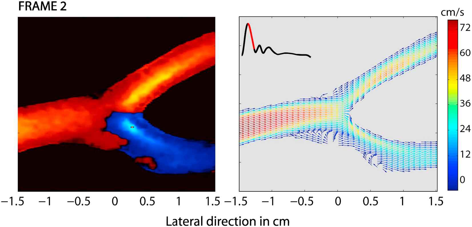
Discussion: Currently used ultrasound imaging modalities have important limitations to assess complex flow in the carotid artery. This complicates the use of these images to extract quantitative data related to flow such as wall shear stress. This virtual ultrasound environment is a powerful tool to assess limitations of currently used ultrasound imaging modalities and to develop new algorithms of upcoming techniques such as 3D ultrasound.
Cite this article
TY  - JOUR
AU  - A. Swillens
AU  - L. Lovstakken
AU  - H. Torp
AU  - P. Segers
PY  - 2008
DA  - 2008/09/15
TI  - P2.21 COMPLEXITY OF 3D CAROTID BIFURCATION BLOOD FLOW PATTERNS IS NOT ADEQUATELY CAPTURED BY CURRENTLY USED ULTRASOUND MODALITIES
JO  - Artery Research
SP  - 111
EP  - 111
VL  - 2
IS  - 3
SN  - 1876-4401
UR  - https://doi.org/10.1016/j.artres.2008.08.387
DO  - 10.1016/j.artres.2008.08.387
ID  - Swillens2008
ER  -