
P3.07 QUANTITATIVE METHOD TO DETERMINE SMOOTH MUSCLE CELL ORIENTATION IN VITAL ARTERIES
- DOI
- 10.1016/j.artres.2012.09.134How to use a DOI?
- Open Access
- This is an open access article distributed under the CC BY-NC license.
Arterial wall tension is actively regulated by smooth muscle cells (SMCs). To study the biomechanical behaviour of the arterial wall, SMC orientation should be accurately quantified. We evaluated the feasibility of a method to quantify SMC orientation in vital, excised arteries imaged by two-photon laser scanning microscopy. Arteries were mounted on micropipettes and pressurised to approximate in-vivo geometry. To enable unambiguous determination of orientation of individual cells, and assuming nuclear orientation to be representative of cell orientation, specimens were stained with a nuclear dye (SYTO13). Images were acquired with increasing imaging depth (Figure). Nuclei were automatically delineated in each image by applying vesselness filtering, a technique that enhances elongated structures. After thresholding, a momentum matrix was calculated for each nucleus, consisting of the sum of pixel to centre-of-gravity distances for each combination of coordinate axes. For each nucleus, the angle θ between its longest axis, as obtained through eigenvalue analysis, and the vessel’s circumferential axis was calculated (Figure). Mean (θm) and SD (θsd) of SMC orientation were calculated by averaging SMC angles over a depth-ROI (Figure) using circular statistics. We analysed image stacks of murine carotid (n=3) and uterine (n=3) arteries. Orientation averaged over all stacks was approximately circumferential (2±5°, mean±SD). These results demonstrate the potential of our approach to quantify SMC orientation in vital arteries, circumventing artefacts associated with histological fixation and sectioning.
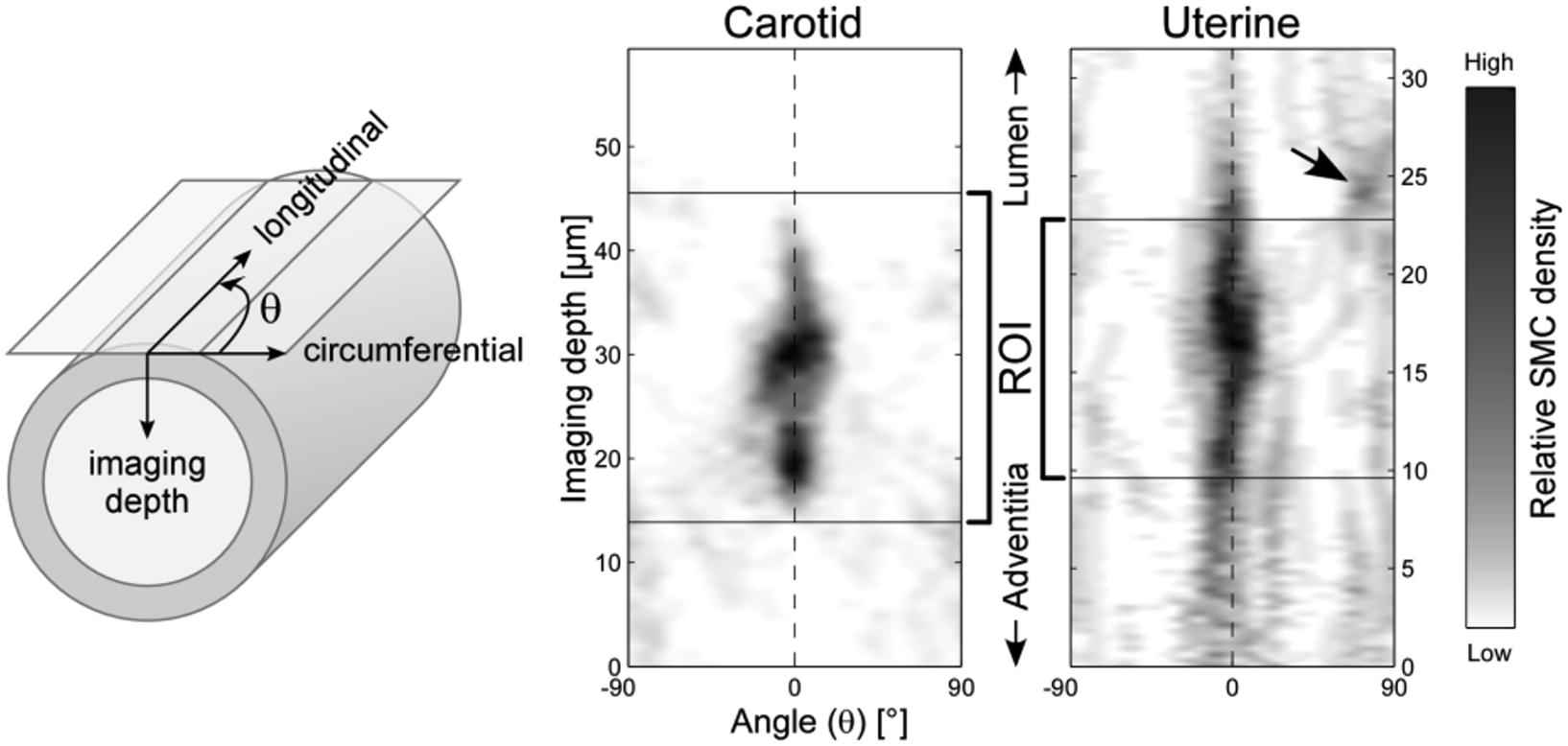
SMC angles as a function of imaging depth for carotid (θm±θsd=−4±25°) and uterine artery (θm±θsd=5±32°). Arrow indicates endothelial cells with elongated nuclei.
Cite this article
TY  - JOUR
AU  - B. Spronck
AU  - J.J. Merken
AU  - W. Kroon
AU  - R.T.A. Megens
AU  - K.D. Reesink
AU  - T. Delhaas
PY  - 2012
DA  - 2012/11/17
TI  - P3.07 QUANTITATIVE METHOD TO DETERMINE SMOOTH MUSCLE CELL ORIENTATION IN VITAL ARTERIES
JO  - Artery Research
SP  - 179
EP  - 179
VL  - 6
IS  - 4
SN  - 1876-4401
UR  - https://doi.org/10.1016/j.artres.2012.09.134
DO  - 10.1016/j.artres.2012.09.134
ID  - Spronck2012
ER  -