
P81 DISARRAY AND REMODELING OF THE RADIAL ARTERY IN WOMEN WITH SPONTANEOUS CORONARY ARTERY DISSECTION: THE FUCHSIA STUDY
- DOI
- 10.1016/j.artres.2017.10.097How to use a DOI?
- Abstract
Background: Spontaneous coronary dissection (SCAD) may represent a manifestation of fibromuscular dysplasia (FMD); thus, preclinical lesions might be found in extracoronary vessels with similar size and wall ultrastructure, such as the radial artery.
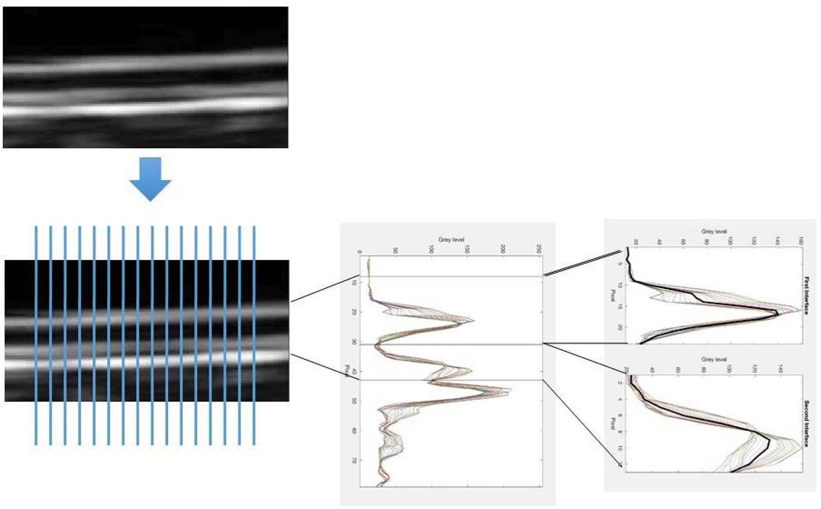
Methods: Two 5′-clips from the left radial artery were obtained by Vevo MD (70 MHz probe, FUJIFILM, VisualSonics). Radial wall showed two echogenic interfaces: the 1st (lumen-media) and the 2nd (media-adventitia). Intima-media (IMT), adventitia (AT), and global thickness (IMAT) and wall cross-sectional area (WCSA) Measured.
Vascular wall disarray was assessed calculating the root mean square error (RMSE) between 20 gray-level profiles crossing the two interfaces and the profile obtained averaging them, normalized for the maximum value of the corresponding mean profile (RMSE/mean).
Results: 5 female SCAD patients and 9 healthy controls (C) were enrolled (age 45±9 vs 45±13years, p = 0.95; BMI 21±3 vs 23±kg/mq, p = 0.22; mean BP 77±5 vs 85±10 mmHg, p = 0.053). 2nd interface peak was reduced in the SCAD group (97±29 130±19, p = 0.04), whereas RMSE/mean was increased (1.89±0.68 vs 0.97±0.30, p = 0.02). Similar values were found for the 1st interface. IMT (0.165±0.031 vs 0.125±0.022mm, p = 0.03), but not AT (0.095±0.020 vs 0.081±0.020mm, p = 0.20) and IMAT (0.260±0.049 vs 0.206±0.030mm, p = 0.053), was significantly higher in SCAD.
Radial internal diameter and wall/lumen ratio were similar: conversely WCSA was increased in SCAD (1.69±0.48 vs 1.07±0.37mm2, p = 0.02).
Conclusions: Radial arteries of SCAD patients were characterized by increased wall thickness. Furthermore, the 2nd echogenic layer exhibited loss of echogenicity and inhomogeneity, features similar to FMD patients.
- Open Access
- This is an open access article distributed under the CC BY-NC license.
Cite this article
TY  - JOUR
AU  - Rosa Maria Bruno
AU  - Nicole Di Lascio
AU  - Abtehale Al Hussaini
AU  - Daniela Guarino
AU  - Saverio Vitali
AU  - Piercarlo Rossi
AU  - Davide Caramella
AU  - Bernardo Cortese
AU  - Francesco Faita
AU  - Stefano Taddei
AU  - Lorenzo Ghiadoni
AU  - David Adlam
PY  - 2017
DA  - 2017/12/06
TI  - P81 DISARRAY AND REMODELING OF THE RADIAL ARTERY IN WOMEN WITH SPONTANEOUS CORONARY ARTERY DISSECTION: THE FUCHSIA STUDY
JO  - Artery Research
SP  - 75
EP  - 76
VL  - 20
IS  - C
SN  - 1876-4401
UR  - https://doi.org/10.1016/j.artres.2017.10.097
DO  - 10.1016/j.artres.2017.10.097
ID  - Bruno2017
ER  -