
Agreement between carotid and radial augmentation index: Does medication status affect the relation?☆
Grant support: JSPS Postdoctoral Fellowships for Research Abroad (JS).
- DOI
- 10.1016/j.artres.2008.03.001How to use a DOI?
- Keywords
- Arterial stiffness; Blood pressure waveform; Applanation tonometry
- Abstract
Central augmentation index (AI) is an index for arterial stiffness and wave reflection, but the measurement requires technical precision. We recently reported that AI obtained directly from radial arterial pressure waveforms (without using the general transfer function) could provide equivalent information to carotid AI in healthy adults. The aim of the present study was to determine whether such association would exist among patients on anti-hypertensive drugs. Forty-six hypertensive patients taking blood pressure lowering medications (62 ± 9 years, mean ± SD) and 78 age-matched apparently healthy adults (60 ± 9 years) were studied. Carotid and radial AI were obtained using arterial applanation tonometry, and radial AI was calculated using the equation [(second peak radial systolic pressure - diastolic pressure)/(first peak radial systolic pressure - diastolic pressure) × 100]. Radial AI was strongly and positively correlated with carotid AI in medicated patients (r = 0.73, P < 0.0001) as well as in healthy controls (r = 0.84, P < 0.0001). The Brand–Altman plot demonstrated that the difference and SD between carotid and radial AI were not different between medicated patients and healthy controls (61.8 ± 7.7 vs 63.0 ± 7.7%). These results suggest that radial AI may be able to provide qualitatively similar information to carotid AI even in patients on antihypertensive medications.
- Copyright
- © 2008 Association for Research into Arterial Structure and Physiology. Published by Elsevier B.V. All rights reserved.
- Open Access
- This is an open access article distributed under the CC BY-NC license.
Background
Central arterial augmentation index (AI) is a useful index of arterial wave reflection that has been directly linked with cardiovascular mortality and morbidity,1,2 but the measurement requires technical precision. We recently described a strong association between carotid AI (a surrogate for central AI) and radial AI obtained directly from pressure waveforms at both locations in healthy subjects.3 However, high-risk patients are often taking anti-hypertensive drugs, which could have differential effects on central (large elastic) and peripheral (muscular) arteries.4,5 If such dissociation exists, it would limit the applicability and utility of the peripherally-measured AI.
Aim
The aim of the present study was to determine whether an association between carotid and radial AI observed in healthy adults would also exist among patients on anti-hypertensive drugs.
Methods
We studied 46 hypertensive patients who were taking blood pressure lowering medications (mean age: 62 ± 9 years) and 78 age-matched healthy controls (60 ± 9 years). All subjects had no apparent overt cardiovascular disease (other than hypertension) as assessed by medical history. The medicated patients used calcium channel blocker (66%), angiotensin receptor blocker (31%), α-blocker (13%), α–β-blocker (3%), angiotensin-converting enzyme inhibitor (3%), diuretics (3%), and vasodilator (i.e., nitric oxide donor, 6%). All subjects gave their written informed consent to participate. This study was reviewed and approved by the local Institutional Review Board.
All measurements were performed after an abstinence of caffeine and a 3-hour fast. After resting in supine position for at least 15 min in a quiet, temperature-controlled room, carotid and radial arterial AI were measured in a random order by two vascular testing devices equipped applanation tonometry probe incorporating an array of micropiezoresistive transducers (VP-2000, Colin Medical Technology; HEM-9010AI, Omron Healthcare) as previously reported.3,6 The characteristic points on the carotid and radial pressure waveform were assessed as previously described.3,7 Heart rate and brachial blood pressure were measured by the vascular testing device (VP-2000, Colin Medical Technology). Mann–Whitney U test was used to determine significant group differences after Kolmogorov–Smirnov and Lilliefors test for normality. Univariate correlation analysis and Bland–Altman plots were used to assess relationships between variables of interest. Forward stepwise multiple-regression analysis was performed to assess independent predictors of radial AI. The effect of the medicated status on the relation between carotid and radial AI was assessed by general linear regression model.
Results
As shown in Table 1, there were no significant differences in height, body weight, and body mass index between the two groups. As expected, systolic and diastolic blood pressures, and carotid AI, were significantly higher in medicated patients than in healthy controls. Radial AI tended to be higher in medicated patients than in healthy controls (P = 0.07). Radial AI was strongly correlated with carotid AI in medicated patients (r = 0.72, P < 0.0001, Fig. 1) as well as in healthy controls (r = 0.84, P < 0.0001, Fig. 1). The Bland–Altman plot demonstrated that the difference and SD between carotid and radial AI in medicated patients were not different from those in healthy controls (62 ± 8 vs. 63 ± 8%, P = 0.40).
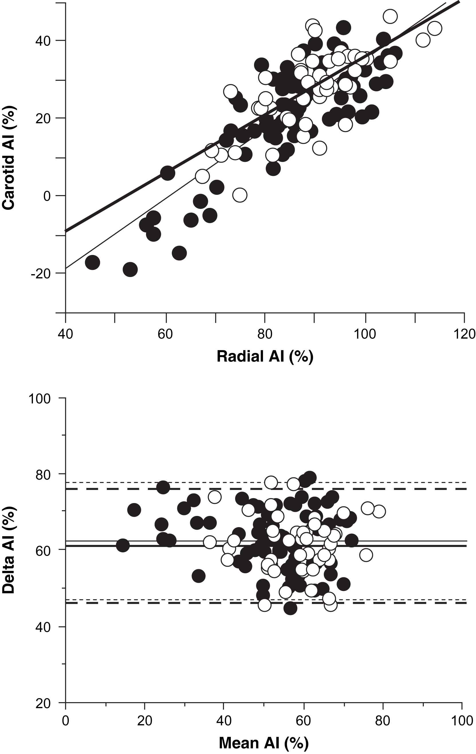
Scatter plots (top) and Bland and Altman’s plots (bottom) between carotid augmentation index (AI) and radial AI in healthy controls (closed circle) and medicated patients (open circle). Thin and thick lines in scatter plots are the regression lines of healthy controls (r = 0.84, P < 0.0001) and of the medicated patients (r = 0.72, P < 0.0001), respectively. Thin (healthy controls) and thick (medicated patients) solid and broken lines in Bland and Altman’s plots showed mean difference (radial AI–carotid AI) and SD, respectively.
| Healthy controls | Medicated patients | |
|---|---|---|
| Male/female | 33/45 | 24/22 |
| Age, years | 60 ± 9 | 62 ± 9 |
| Height, cm | 160 ± 9 | 161 ± 8 |
| Weight, kg | 59 ± 10 | 63 ± 10 |
| BMI, kg/m2 | 23.1 ± 2.9 | 24.1 ± 2.9 |
| Heart rate, bpm | 62 ± 9 | 61 ± 7 |
| Systolic BP, mmHg | 121 ± 10 | 135 ± 20* |
| Diastolic BP, mmHg | 74 ± 8 | 82 ± 13* |
| Carotid AI, % | 21.5 ± 14.0 | 27.3 ± 10.6* |
| Radial AI, % | 84.5 ± 12.9 | 89.1 ± 10.3 |
Data are mean ± SD.
P < 0.05 vs. healthy controls.
BMI = body mass index, BP = blood pressure.
Physical characteristics
In healthy controls, radial AI was significantly correlated with age (r = 0.61), sex (r = 0.59), height (r = −0.60), weight (r = −0.48), and heart rate (r = −0.40) as well as carotid AI. A forward stepwise regression analysis revealed that carotid AI (beta = 0.58), age (beta = 0.17), sex (beta = 0.16), and heart rate (beta = −0.16) were entered the model as independent predictors of radial AI (adjusted multiple R2 = 0.73, P < 0.0001). Similarly, the medicated patients showed significant relationship of radial AI to heart rate (r = −0.54), age (r = 0.33), sex (r = 0.28), and height (r = −0.23) as well as carotid AI. A forward stepwise regression analysis revealed that carotid AI (beta = 0.58) and heart rate (beta = −0.30) were entered the model as independent predictors of radial AI in the medicated patients (adjusted multiple R2 = 0.59, P < 0.0001). General linear regression model revealed that radial AI was a significant independent predictor for carotid AI in the pooled subjects and that the medicated status was not entered into the model, suggesting that the medication status did not interfere with the slope of the relation between carotid and radial AI.
Discussion
Considering the increasing emphasis placed on primary prevention of cardiovascular disease, the development of noninvasive techniques to screen high-risk patients is clearly meaningful. Our results suggest that radial AI, which is easier to measure and implement, may be able to provide qualitatively similar information to carotid AI even in patients on anti-hypertensive medications. Our findings would extend our previous findings in healthy humans3 to medicated hypertensive population.
As vasoactive agents can affect large and/or small arteries’ vascular tone differentially,4,5 a question can be raised as to why a constant association between carotid and radial AI was observed regardless of the medication status. Augmentation index incorporates the magnitude as well as the timing of the incident wave from the heart and the reflected wave from the periphery.8 Since major paths of pressure wave (e.g., aorta) are common to both carotid and radial AI, acute and/or chronic influences of vasoactive agents might be expected to be similar on both carotid and radial AI. In this context, we have previously reported that acute changes in radial AI produced by sympathetic nervous activity stimulation were closely associated with the corresponding changes in carotid AI.3
In the present study, all subjects were Japanese, and majority of them were taking calcium channel blocker and/or ARB. We recognize that the approach for treating hypertension are different among countries (i.e., Japan vs. US)9,10 and that the results might be different when similar studies are conducted in a different country. Unfortunately, our sample size was too small to analyze whether the type of drug may affect the relation between carotid and radial AI. Further study to address this issue is warranted.
Acknowledgement
The autonomic device tested for the present study was provided by the Omron Health Care Corporation, Kyoto, Japan.
References
Cite this article
TY  - JOUR
AU  - Jun Sugawara
AU  - Hidehiko Komine
AU  - Koichiro Hayashi
AU  - Mutsuko Yoshizawa
AU  - Takashi Yokoi
AU  - Seiji Maeda
AU  - Hirofumi Tanaka
PY  - 2008
DA  - 2008/04/28
TI  - Agreement between carotid and radial augmentation index: Does medication status affect the relation?☆
JO  - Artery Research
SP  - 74
EP  - 76
VL  - 2
IS  - 2
SN  - 1876-4401
UR  - https://doi.org/10.1016/j.artres.2008.03.001
DO  - 10.1016/j.artres.2008.03.001
ID  - Sugawara2008
ER  -