
An fNIRS Study of Brain State during Letter and Category Fluency Tasks
- DOI
- 10.2991/jrnal.k.190220.003How to use a DOI?
- Keywords
- Verbal fluency task; letter fluency task; category fluency task; fNIRS; brain activity
- Abstract
Verbal Fluency Tasks (VFT) can be categorized into the Letter Fluency Task (LFT) that evaluates the ability to generate words starting with a particular letter, and the Category Fluency Task (CFT) that involves recall of words that belong to semantic categories. In this study, we investigated the differences in brain activity during LFT and CFT in healthy subjects. Brain activity in the frontal and the temporal region was measured using functional near-infrared spectroscopy with 40 channels. We observed more prominent brain activity when performing LFT than CFT.
- Copyright
- © 2019 The Authors. Published by Atlantis Press SARL.
- Open Access
- This is an open access article distributed under the CC BY-NC 4.0 license (http://creativecommons.org/licenses/by-nc/4.0/).
1. INTRODUCTION
Functional Near-infrared Spectroscopy (fNIRS) is a technique that measures metabolic changes in hemoglobin oxygen in vivo, using near-infrared light. fNIRS detects changes in concentration of oxy-hemoglobin, deoxy-hemoglobin, total hemoglobin, and measures alteration in cerebral blood flow caused by neural activity [1,2]. Brain function measurement using fNIRS is noninvasive, has excellent temporal resolution, and is performed under routine conditions. In recent times, fNIRS as a tool in advanced medicine is attracting attention. fNIRS examination using Verbal Fluency Task (VFT) is widely being used in diagnosis of depression symptoms. VFT is a task that requires to generate as many words within a category or starting with a given letter as possible within a time limit. In the Letter Fluency Task (LFT), words are recalled from particular letter. In the Category Fluency Task (CFT), words are recalled from semantic categories. The characteristics of brain activity in VFT are different in depression, bipolar disorder and schizophrenia, therefore, diverse activating tasks are used for different disorders. It has been pointed out that VFT tests not only the semantic memory related to vocabulary but also many cognitive functions such as effective vocabulary search power, information processing speed, and executive function [3]. There are several studies on brain activity differences in LFT and CFT of healthy subjects and patients with a mental illness [4–8]. However, only a few studies compare LFT and CFT as such. In this study, brain activities during LFT and CFT were compared in healthy subjects. We hypothesized a difference in brain activity between LFT and CFT and therefore, the purpose of the current study was to investigate the brain status of the two versions of the VFT over a wide area of the frontal and bilateral temporal brain regions using 40 channels of fNIRS.
2. MATERIALS AND METHODS
2.1. Participants
Healthy subjects (average age 22.35 ± 0.93, 17 males, three females, right-handed) were recruited for this study. All the participants performed LFT and CFT, and brain activity during each task was measured using fNIRS. This study was approved by the Ethics Committee of the Doshisha University.
2.2. Experiment Procedure
In the experiment, a block-design VFT (letter and category version) was performed. The experimental design is shown in Figure 1. It consisted of 60 s of pre-task rest followed by 60 s of task period. Participants were initially instructed to watch a cross at the center of the computer screen during the experiment. The task section began with three tasks (a, ka, shi/animals, fruits, sports). This was explained to the participants by the computer as audible instructions. The content of the task was determined from the frequency of use of letters and previous studies [8,9]. During LFT, participants were instructed to generate as many words as possible starting with the displayed letters, while during CFT, participants were instructed to generate as many words which would fit in the presented category. The two tasks were done consecutively and the participants were explained about the order at the beginning of the experiment. The total number of correct words generated during the task was recorded as an indicator of VFT performance.
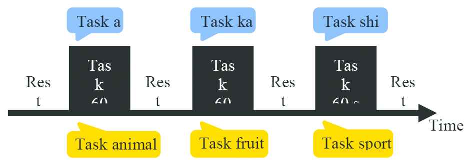
Experimental design.
2.3. NIRS Instrument
In this study, a 40-channel fNIRS device (LABNIRS, Shimadzu Corporation, Kyoto) was used. The distance between the irradiation probe and the light receiving probe was set to 3.0 cm. To minimize the artifacts, the participants were instructed to avoid voluntary movements. The experimental set-up is shown in Figure 2. The sampling frequency was set at 37.037 Hz. After data acquisition, three-dimensional (3D) co-ordinates of each light source and detector were measured using a 3D digitizer (FASTRAK, Polhemus, Vermont, USA). Subsequently, these were input to the Statistical Parametric Mapping (SPM)-fNIRS toolbox for spatial registration, and the layout of each channel and Montreal Neurological Institute co-ordinates was created.
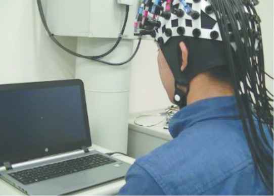
Experimental set-up.
2.4. Data Processing
Functional near-infrared spectroscopy data was band-pass filtered using 0.01–0.1 Hz. fNIRS data from nine participants were excluded due to a variation of 0.01 mM * cm in 0.1 s. fNIRS data was band-stop filtered (physiological noise removal) using 0.12–0.35 Hz, and with 0.7–1.5 Hz after artifact correction based on moving standard deviation and spline interpolation. Next, trend removal was performed using a high pass filter based on the discrete cosine transform set. General linear model analysis was used for extraction of the active regions in the brain. Using the hemodynamic response function, a blood flow alteration model was created as shown in Figure 3. Statistical significance in brain activity between the two groups was examined using the regression coefficient obtained by regression analysis of blood flow alteration model and measurement data. All the calculations and modeling was done using the SPM-fNIRS toolbox.
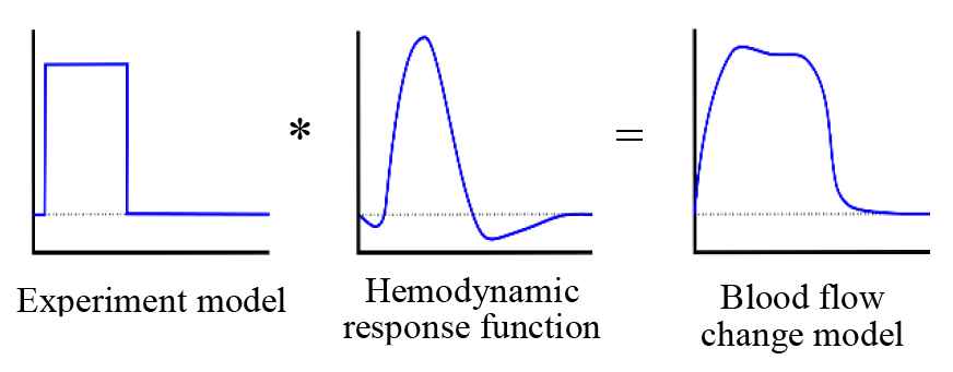
Creation of blood flow alteration model.
3. RESULTS
Brain activation analysis was performed with 11 fNIRS data. Regions with maximum activity for LFT and CFT are represented in Tables 1 and 2, respectively. Group analysis results are shown in Tables 3 and 4. Brain activity observed during LFT performance was higher than during CFT.
| Region | Number of subjects |
|---|---|
| Precentral L | 10 |
| Precentral R | 10 |
| Frontal Sup L | 8 |
| Frontal Sup R | 8 |
| Frontal Mid L | 10 |
| Frontal Mid R | 8 |
| Frontal Mid Orb L | 7 |
| Frontal Inf L | 10 |
| Frontal Inf R | 10 |
| Postcentral L | 9 |
| Postcentral R | 9 |
| SupraMarginal L | 8 |
| SupraMarginal R | 9 |
| Angular R | 7 |
| Temporal Sup L | 8 |
| Temporal Sup R | 8 |
| Temporal Mid L | 8 |
| Temporal Mid R | 6 |
| Temporal Inf L | 7 |
Activated brain region (LFT) (FWE, p < 0.05)
| Region | Number of subjects |
|---|---|
| Precentral L | 7 |
| Precentral R | 7 |
| Frontal Sup L | 7 |
| Frontal Sup R | 6 |
| Frontal Mid L | 7 |
| Frontal Mid R | 7 |
| Frontal Inf L | 7 |
| Frontal Inf R | 7 |
| Postcentral L | 7 |
| Postcentral R | 6 |
| SupraMarginal L | 6 |
| SupraMarginal R | 6 |
| Temporal Sup L | 6 |
| Temporal Mid L | 6 |
| Temporal Inf L | 6 |
Activated brain region (CFT) (FWE, p < 0.05)
| Region |
|---|
| Rolandic Oper L |
| Temporal Sup R |
| Frontal Inf Tri L |
| Frontal Inf Tri R |
Group analysis result (LFT) (FWE, p < 0.05)
| Region |
|---|
| Frontal Inf Tri R |
| Caudate R |
Group analysis result (CFT) (FWE, p < 0.05)
4. DISCUSSION
In this study, brain activation was investigated based on the change in hemoglobin concentration measured during VFT performance in healthy subjects. An extensive activation in the frontal and temporal brain regions including Broca’s areas was observed during the VFT performance. In particular, LFT augmented the brain activation compared with CFT. During CFT, participants would search for words by utilizing the concept of associations such as already existing vehicles. On the other hand, in LFT, words were searched from the notion of phonology which is rarely used in daily life. In other words, participants suppress the recall of related words and perform complex word searches [10]. The results of this study suggest that LFT requires more activation of brain regions than CFT. As shown in Tables 1 and 2, four regions are activated exclusively during LFT. The regions include Angular Gyrus R (R-ANG), Middle Temporal Gyrus R (R-MTG), Superior Temporal Gyrus R (R-STG), and Orbital Part of Inferior Frontal Gyrus L (L-ORB) as shown in Figure 4. Amongst these four regions, ANG is located at the junction of the occipital, temporal, and parietal regions and is regarded as an important interface for communicating and integrating information with various brain regions [11,12]. Several studies have associated ANG with memory, meaning, memory consciousness, default mode network [13]. MTG, on the other hand, is activated with a more difficult phonological task and plays an important role in controlling semantic search [14]. Co-activation of ANG and MTG observed during LFT suggests their role in word searches that suppresses related word remembrance.
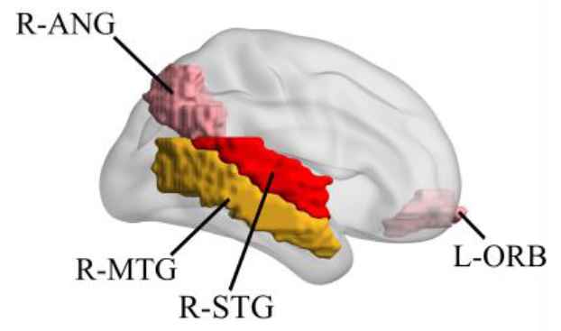
Brain regions activated only during LFT.
5. CONCLUSION
Currently, several ongoing studies are investigating the difference in brain activity between LFT and CFT in healthy subjects and patients with a mental illness. However, there are only a few studies that compare LFT and CFT as such. Therefore, in this study, the brain activities during LFT and CFT were compared only in healthy subjects. The results from 11 healthy subjects established an increase in LFT activation compared to CFT. Our results suggest that co-activation of ANG and MTG makes it possible to search words with related word recall suppressed.
Authors Introduction
Ms. Akane Onishi
 Akane Onishi is currently studying toward the bachelor’s degree in Engineering at Faculty of Life and Medical Sciences, Doshisha University, Kyoto, Japan. Her research interests include cognitive neuroscience, with a particular focus on neuroimaging using functional near-infrared spectroscopy.
Dr. Hiroshi Furutani
 Hiroshi Furutani received PhD of Science from Kyoto University in 1981. He is currently working in Innovative Computing Research Center at Doshisha University as researcher. His research interests include bio-informatics, evolutionary computation and quantum computing.
Dr. Tomoyuki Hiroyasu
 Tomoyuki Hiroyasu received the Doctor of Engineering from Waseda University in 1998. He has been a Professor of Life and Medical Sciences, Doshisha University, since 2008. His research interests include the development of novel evolutionary algorithms, medical image processing, medical information systems, parallel computing, Grid, web systems, and Intelligent systems. Prof. Hiroyasu is a member of IEEE, JSME, JSCES, IPSJ, SICE, and IEICE.
Dr. Satoru Hiwa
 Satoru Hiwa received PhD in Engineering from Doshisha University. He is currently working in Faculty of Life and Medical Sciences at Doshisha University, Kyoto, Japan, as an Assistant Professor. His current research interests include human brain mapping, noninvasive neuroimaging and computational intelligence.
REFERENCES
Cite this article
TY  - JOUR
AU  - Akane Onishi
AU  - Hiroshi Furutani
AU  - Tomoyuki Hiroyasu
AU  - Satoru Hiwa
PY  - 2019
DA  - 2019/03/30
TI  - An fNIRS Study of Brain State during Letter and Category Fluency Tasks
JO  - Journal of Robotics, Networking and Artificial Life
SP  - 228
EP  - 231
VL  - 5
IS  - 4
SN  - 2352-6386
UR  - https://doi.org/10.2991/jrnal.k.190220.003
DO  - 10.2991/jrnal.k.190220.003
ID  - Onishi2019
ER  -