
A Study for the Realization of Online Magnetoencephalography using the Spatio-Spectral Decomposition Algorithms
- DOI
- 10.2991/jrnal.k.210922.002How to use a DOI?
- Keywords
- Magnetoencephalography; spatio-spectral decomposition; Morlet wavelet transform; neurofeedback
- Abstract
Neurofeedback systems have been found to be effective in the clinical rehabilitation of paralysis. However, most systems exist only for use with electroencephalography, which is cumbersome to apply to patients and has lower spatial resolution than Magnetoencephalography (MEG). Furthermore, the best practices for neural data feature extraction and feature selection are not well established. The inclusion of the best performing feature extraction algorithms is critical to the development of clinical neurofeedback systems. Using simultaneously collected MEG and accelerometer data before and during 10 spontaneous finger movements, we performed an in-depth comparison of the Spatio-Spectral Decomposition (SSD) algorithms for their individual abilities to isolate movement-relevant features in brain activity. Having restricted raw data to that from sensorimotor rhythm frequencies in select MEG sensors over sensorimotor cortex, we compared SSD components using: (1) 2D topographies, (2) activations over time, (3) and correlations with accelerometer data at both 0 and 60 ms time delays. We will discuss these results and suggestions for application to neurofeedback systems. In particular, we will present detailed visualizations of SSD results and discuss potential strategies and pitfalls for feature selection.
- Copyright
- © 2021 The Authors. Published by Atlantis Press International B.V.
- Open Access
- This is an open access article distributed under the CC BY-NC 4.0 license (http://creativecommons.org/licenses/by-nc/4.0/).
1. INTRODUCTION
For diseases that affect brain function, such as strokes, immediate treatment with medication and surgery is important, but post-onset rehabilitation also plays a critical role in the wellbeing of patients.
One of the techniques used for non-invasive brain function evaluation is Magnetoencephalography (MEG) [1,2]. MEG has high temporal resolution as well as high spatial resolution, and it is commonly used clinically for epilepsy diagnosis and rehabilitation [3].
In the past, electroencephalography was often used for the real time feedback system [4,5]. Real-time feedback system using MEG has been reported in the field of Brain–Machine Interface [6]. However, rehabilitation of the real time feedback system using MEG has not been established.
However, without the ability to monitor a patient’s relevant brain activity in real time during rehabilitative exercises, the most efficient rehabilitation cannot occur.
The purpose of this study was to evaluate the use of Spatio-Spectral Decomposition (SSD) of real-time MEG data during spontaneous movement [7–10]. Performance was evaluated by comparing SSD results to the results from a standard analysis technique, Independent Components Analysis (ICA). SSD completed decomposition faster than did ICA, with the SSD analysis completed about 270 times faster in the preliminary experiment. Because processing speed is critical to neurofeedback systems, we present our investigation of the SSD components and conclude with suggestions for feature selection.
2. EXPERIMENT
The data were collected using a full-head 306-channel magnetoencephalograph (Vectorview, Elekta-Neuromag, Helsinki, Finland) at a sampling frequency of 1000 [Hz]. To reduce power supply noise and other interference, measurements were taken inside a magnetic field shield room [1 (kHz) shielding rate 55.2 (dB)]. The participant was a healthy person who attached an acceleration sensor to the middle finger of his left hand and performed self-initiated spontaneous flexion (i.e., bending) and extension (i.e., relaxing) movements of the indicated finger as shown in Figure 1 (hereinafter referred to as “the task”). The accelerometer data indicated the start and duration of flexion and extension.
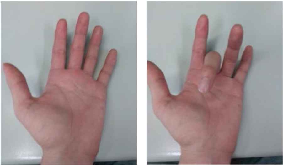
An example of finger extension (i.e., relaxing; left image) and flexion (i.e., bending; right image).
3. ANALYSIS METHOD
After collecting the raw MEG data from all 306 channels, we selected 26 gradiometers corresponding to the right Sensorimotor Cortex and performed ICA and SSD analyses [11,12], which each identified components within the largest cited frequency band for Sensorimotor Rhythm (SMR) [13], 8–30 (Hz).
We performed SSD analysis from 40 to 100 s of data collection (the period of task performance). SSD extracted oscillations with optimized signal-noise ratio (SNR) by maximizing the power from 8 to 30 Hz while simultaneously suppressing the flanking frequency bins. Then we performed a Morlet wavelet transform on the components in order to examine frequency power over time. As seen in Figure 2, high amplitude artifactual data was recorded between 40 and 50 s. To isolate SSD performance during non-artifact contaminated neural activity and to simplify our preliminary analyses, we restricted data analysis to the 20 s from 50 to 70 s. There were 10 tasks completed at irregular intervals over this time period.
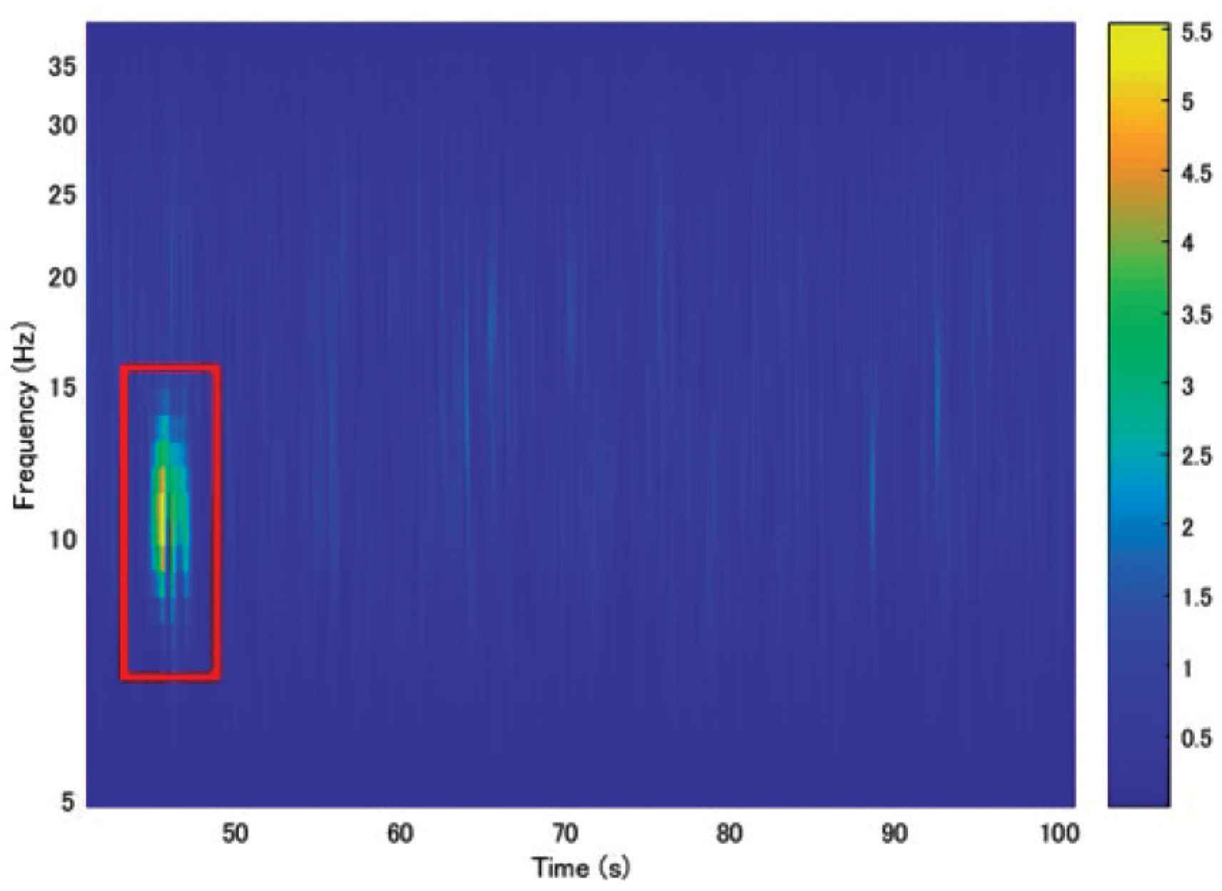
The Morlet wavelet transform (5–40 Hz) of the first SSD component from 40 to 100 s.
4. RESULTS
Following the results of topography (Figure 3), we investigated the first, second, third, and 10th SSD components (ranked from highest to lowest eigenvalue).
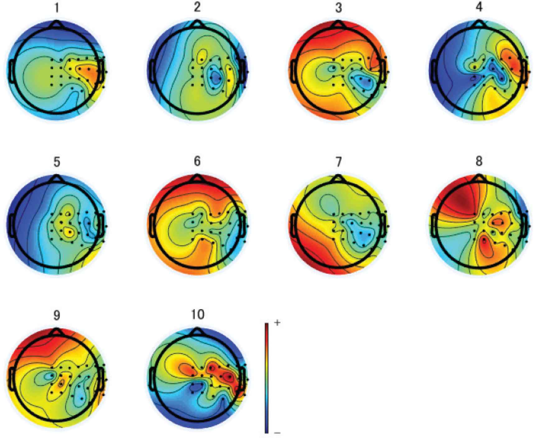
The 2D topographies of SSD components 1 through 10.
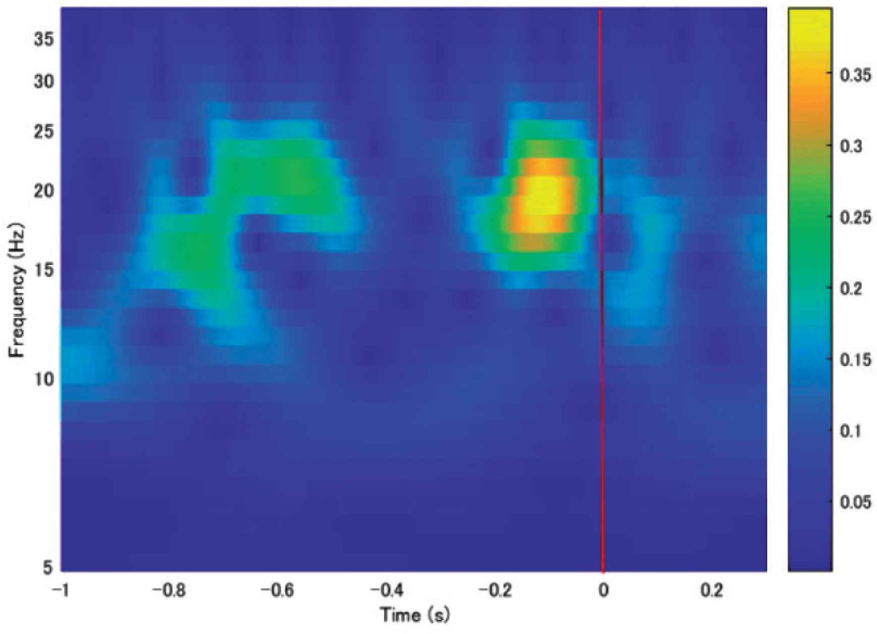
The across-task averaged third and fourth SSD components had higher SMR band power immediately preceding the task. As shown in Figures 4–7, wavelet analysis was performed on task-locked averaged components, indicating average brain activity in the final second before flexion start. The red line in the figure is the flexion start point. The third (Figure 6) and 10th (Figure 7) components each showed a decrease in power around 10–25 Hz immediately following task execution (Black frame), and this decrease in power was followed by a subsequent increase in power from around 10–15 Hz (Red frame).
The Morlet wavelet transform (5–40 Hz) of the task-averaged first SSD component.
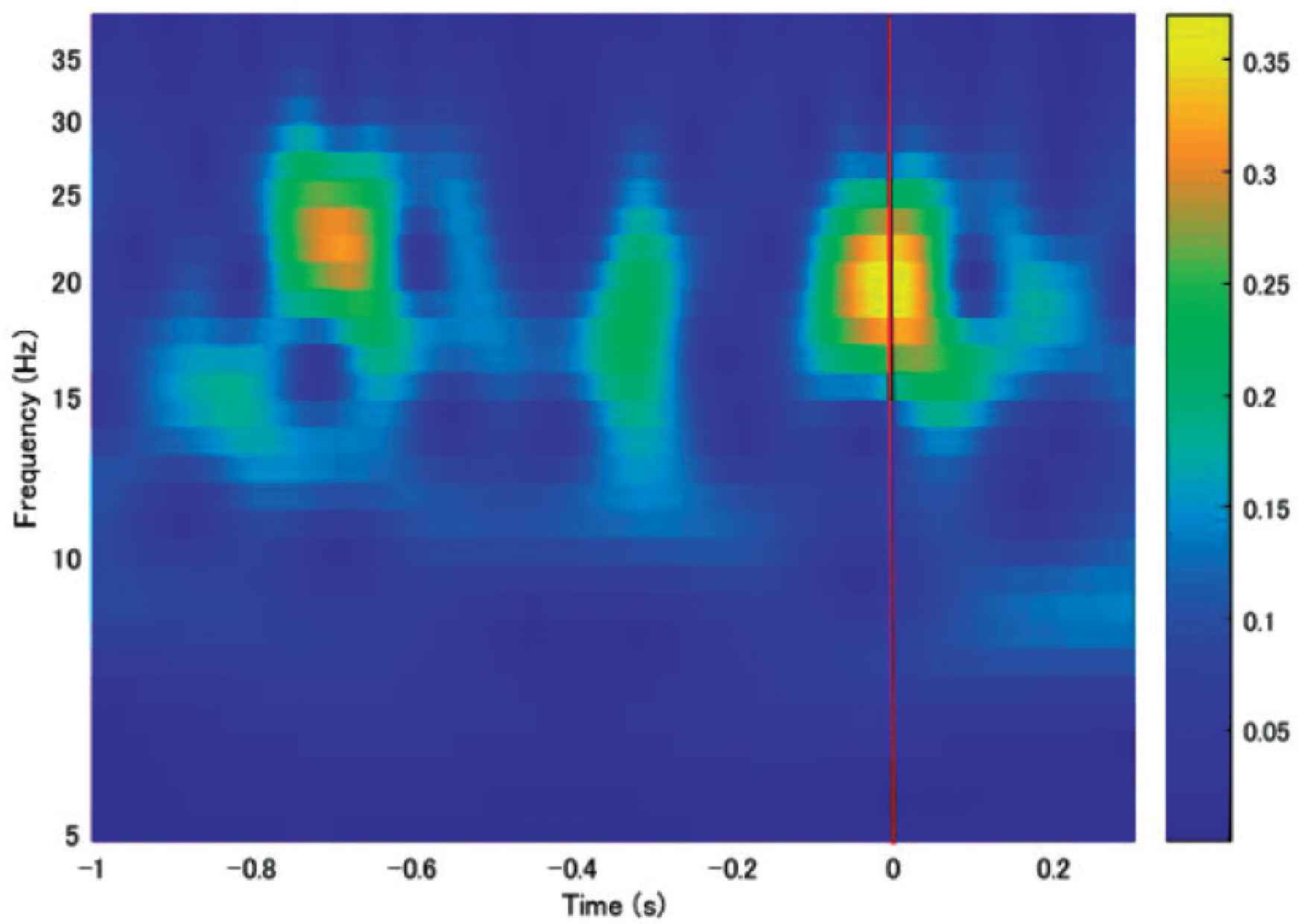
The Morlet wavelet transform (5–40 Hz) of the task-averaged second SSD component.
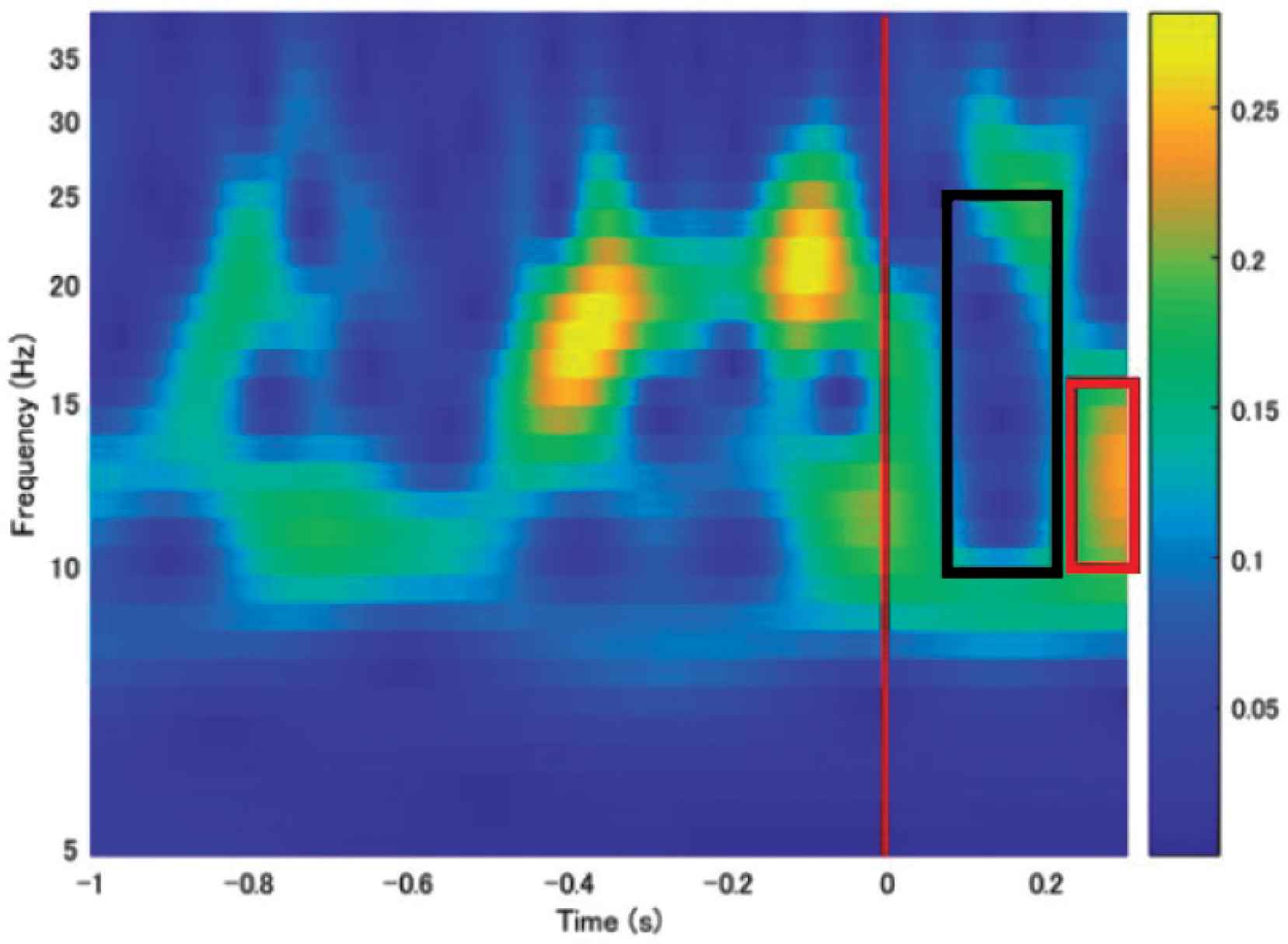
The Morlet wavelet transform (5–40 Hz) of the task-averaged third SSD component.
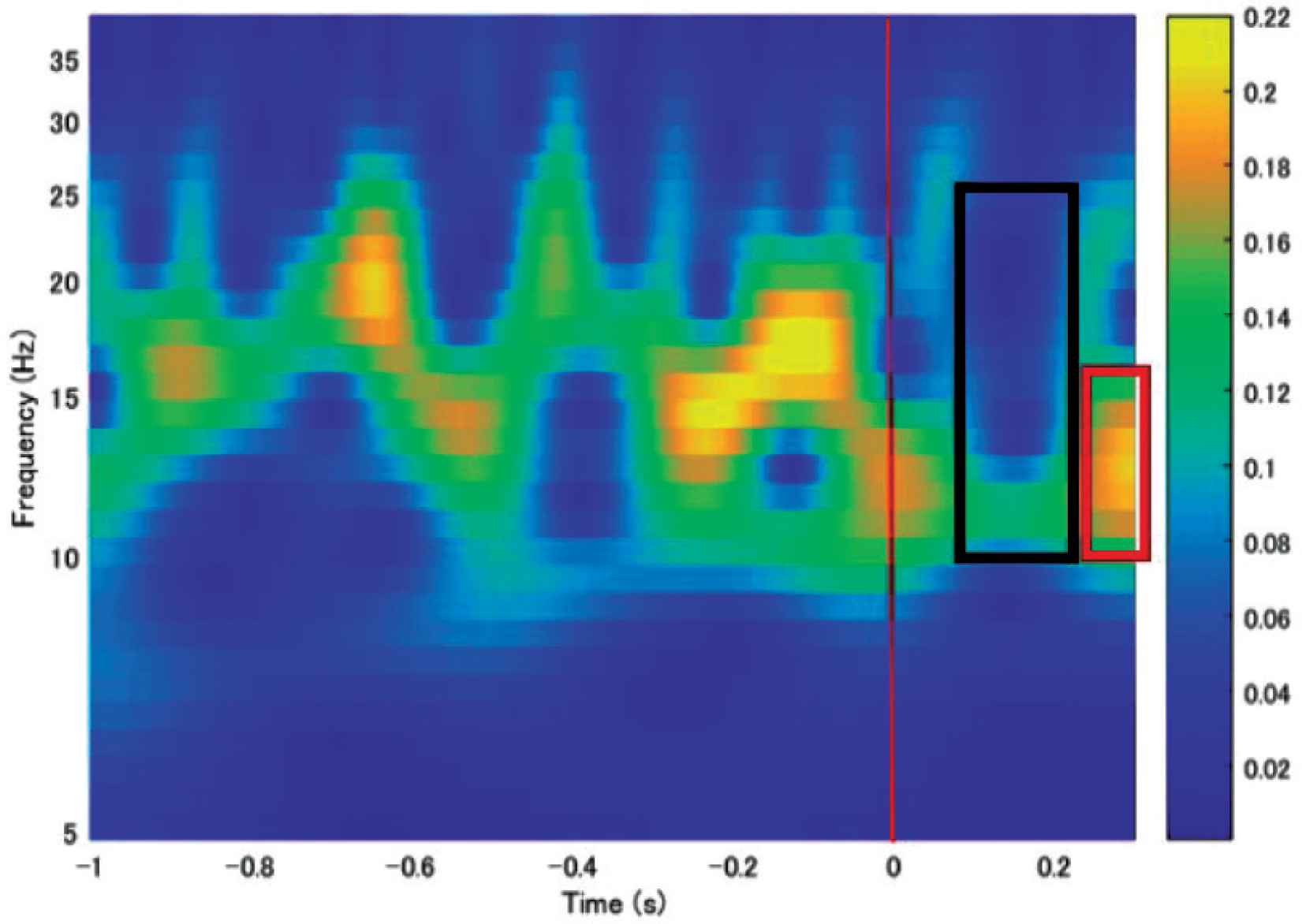
The Morlet wavelet transform (5–40 Hz) of the task-averaged 10th SSD component.
As shown in Table 1, the third component had the highest correlation with the accelerometer data, followed by the second and fourth components which had small but likely still relevant correlations with the accelerometer data.
| 60 (ms) | |
|---|---|
| First SSD | –0.037 |
| Second SSD | –0.28 |
| Third SSD | 0.35 |
| Fourth SSD | –0.24 |
| Fifth SSD | –0.13 |
The correlations between the accelerometer data and the first five SSD components
5. CONCLUSION
In this paper, we performed offline MEG data analysis during spontaneous flexion and extension of the middle finger of the left hand to investigate the use of SSD in an online, real-time MEG-based neurofeedback system.
There are several key points to take away regarding the automation of feature selection in this context. As shown in Table 1, the third SSD component had the highest correlation with the accelerometer data out of the first five components (i.e., the components with the strongest eigenvalues or largest SNRs), although this correlation was still under 0.5. The wavelet analysis revealed Event-related Desynchronization (ERD) and Event-related Synchronization (ERS) in both the third (Figure 6) and 10th (Figure 7) SSD components. Therefore, it is not possible to simply select the first SSD component or the first few SSD components and assume correspondence with the task, nor is it possible to select relevant components based solely on a high-threshold correlation with behavioral data. From previous study, we have also found that selection of the component with the highest SMR power is ineffective due to contamination by muscular artifacts. This analysis leads us to conclude that domain-informed feature selection, in this case using ERD and ERS characteristics, should be the basis for the ideal neurofeedback system (i.e., a system that allows for maximum flexibility and change in brain activity during rehabilitation sessions).
Future work will involve analyzing the correlations between more of the SSD components (e.g., 6–10) and the accelerometer data at various delays. Furthermore, we will investigate the usefulness of baseline correction in component selection and of logarithmically scaled components as features in unsupervised learning.
CONFLICTS OF INTEREST
The authors declare they have no conflicts of interest.
AUTHORS INTRODUCTION
Mr. Kazuhiro Yagi
 He is a clinical laboratory technician at Junwakai memorial hospital in Japan. He is currently a Doctoral course student in Interdisciplinary Graduate School of Agriculture and Engineering, University of Miyazaki. His main research interest is online Magnetoencephalography (MEG).
Mr. Yuta Shibahara
 He received the B.E. degree from Miyazaki University in 2020. He is currently a Master course student in Graduate School of Engineering, University of Miyazaki. His main research interest is online Magnetoencephalography (MEG).
Dr. Lindsey Tate
 She received a B.A. in Psychology (Neuroscience) from St. Edward’s University in Austin, TX, USA in 2013. In 2016 and 2018, respectively, she received her M.S. and PhD in Cognitive Psychology from the University of Oklahoma, where she conducted perceptual and behavioral neuroscience research using EEG and MEG. Now she works as a researcher in the Faculty of Engineering at the University of Miyazaki. Her main research interests are applying machine learning and other algorithms to real-time non-invasive physiological data for use in clinical and non-clinical biofeedback systems.
Dr. Keiko Sakurai
 She received B.E. and M.E. degrees from Kyushu Institute of Technology in 2005 and 2007. From 2007 to 2010, she was in charge of the design of the drive for PC at Pioneer Corporation in Japan. She was engaged in research on both high noise reduction and energy saving. She graduated Doctor course at Interdisciplinary Graduate School of Agriculture and Engineering in University of Miyazaki. Now she works as a researcher in Faculty of Engineering at University of Miyazaki. Her main research interests are biological signal measurement systems, especially gaze estimation system.
Prof. Hiroki Tamura
 He received the B.E. and M.E. degree from Miyazaki University in 1998 and 2000, respectively. From 2000 to 2001, he was an Engineer in Asahi Kasei Corporation, Japan. In 2001, he joined Toyama University, Toyama, Japan, where he was a Technical Official in the Department of Intellectual Information Systems. In 2006, he joined Miyazaki University, Miyazaki, Japan, where he was an Assistant Professor in the Department of Electrical and Electronic Engineering. Since 2015, he is currently a Professor in the Department of Environmental Robotics. His main research interests are Neural Networks and Optimization Problems. In recent years, he has had interest in Biomedical Signal Processing using Soft Computing.
REFERENCES
Cite this article
TY  - JOUR
AU  - Kazuhiro Yagi
AU  - Yuta Shibahara
AU  - Lindsey Tate
AU  - Keiko Sakurai
AU  - Hiroki Tamura
PY  - 2021
DA  - 2021/10/09
TI  - A Study for the Realization of Online Magnetoencephalography using the Spatio-Spectral Decomposition Algorithms
JO  - Journal of Robotics, Networking and Artificial Life
SP  - 161
EP  - 164
VL  - 8
IS  - 3
SN  - 2352-6386
UR  - https://doi.org/10.2991/jrnal.k.210922.002
DO  - 10.2991/jrnal.k.210922.002
ID  - Yagi2021
ER  -