
A case report: How a failure of reversal in arterial flow during development lead to the hypoplasia of a posterior cerebral artery and a resulting incomplete Circle of Willis
- DOI
- 10.1016/j.artres.2016.08.001How to use a DOI?
- Copyright
- © 2016 Association for Research into Arterial Structure and Physiology. Published by Elsevier B.V. All rights reserved.
- Open Access
- This is an open access article distributed under the CC BY-NC license.
Anatomical variations of the Circle of Willis have been well documented, however, they are rarely photographically documented .1–3 This anatomical variation was discovered during the dissection class of the undergraduate anatomy module within the medicine curriculum of Queen’s University Belfast. This specimen displays a physiological failure of the cerebral arteries to undergo reverse flow during development, this lead to the hypoplasia of a right posterior cerebral artery and a resulting incomplete Circle of Willis.
This case reports a malformation of a right posterior cerebral artery, as shown in Fig. 1, found in an 86-year-old female cadaver. An in depth overview of the anatomy of the posterior cerebellar artery and its embryonic development provided.
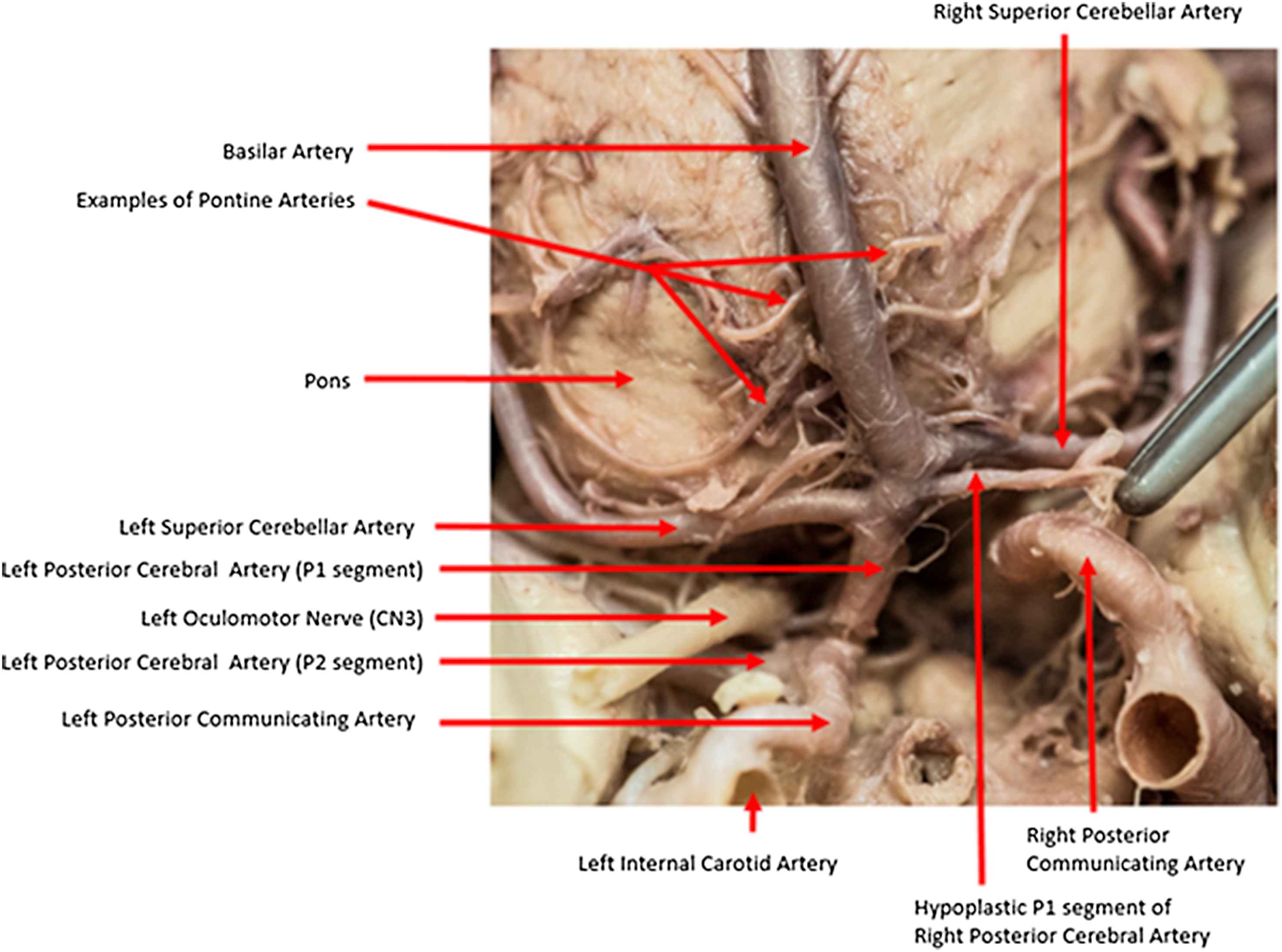
Inferior image of the hypoplastic right posterior communicating artery.
The Circle of Willis is located on the inferior surface of the cerebrum and is antero-superior to the cerebellum. It supplies these regions via the anterior (ACA), middle (MCA) and posterior cerebral arteries (PCA). The anterior segment of the Circle of Willis is comprised of the ACA and MCA, the major terminal branches of the internal carotid artery (ICA).
The posterior segment originates from the basilar artery and terminates as the posterior cerebral artery (PCA). The anterior and posterior segments are linked by two posterior communicating arteries (PcomA), forming an anastomotic Circle as demonstrated in Fig. 2a.
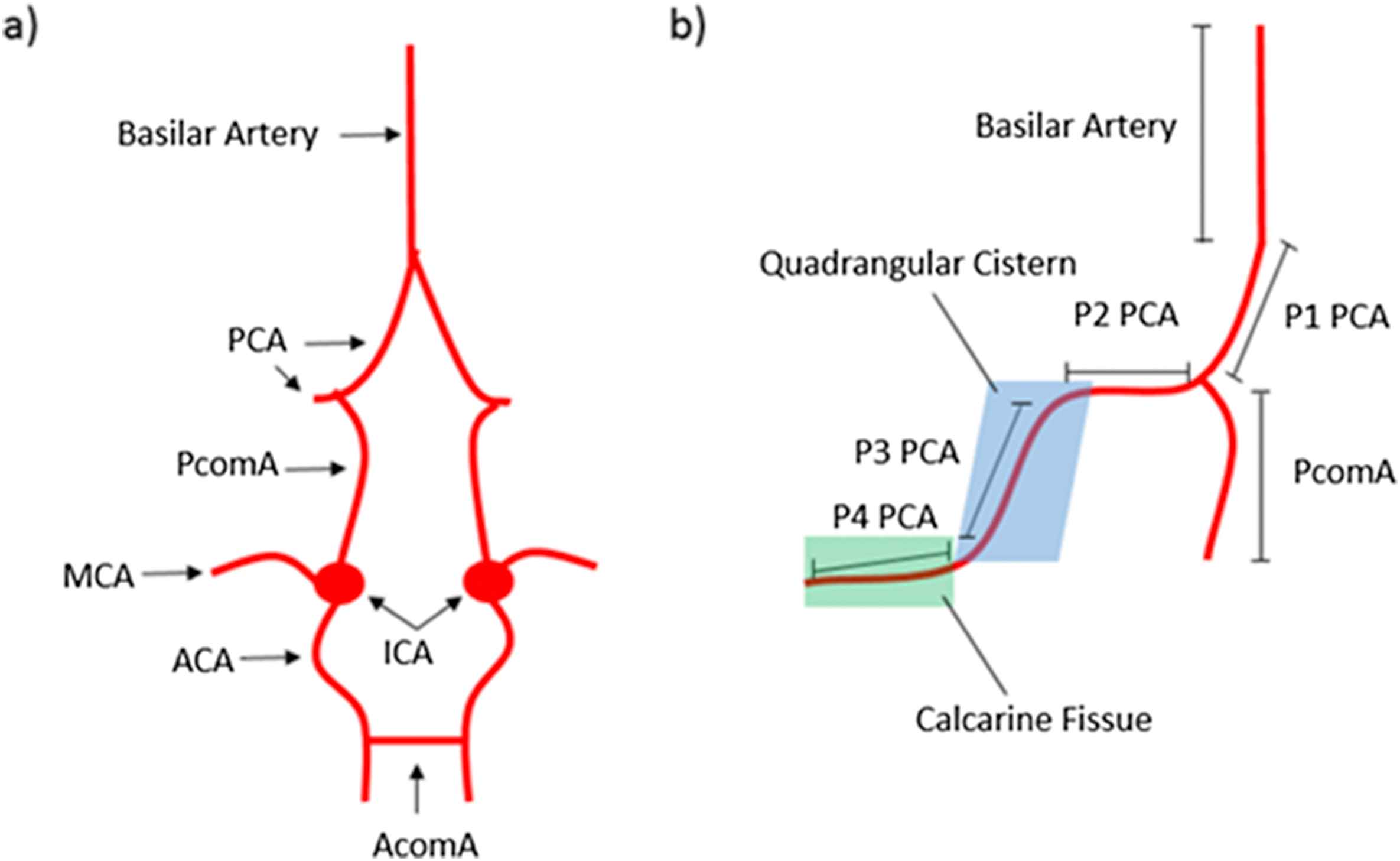
a) Diagram of typical Circle of Willis b) Diagram of the segments of the PCA.
The PCA can be divided into 4 segments. The P1 segment ends at the PcomA and the P2 segment ends on entry to the quadrigeminal cistern, superior to the cerebellum. P3 is located in the quadrigeminal cistern and P4 is the cortical segment located in the calcarine fissure, as illustrated in Fig. 2b.
For us, an understanding of the embryological development of the PCA and the PcomA will be key to understanding the malformation seen in this case report.
By day 23 of embryonic development nutrition supplied by amniotic diffusion is no longer enough to allow the neural tube and noticord to keep developing, and the dorsal aortas take over. Soon these longitudinal vessels running alongside the neural crest can also no longer supply the needs of the growing embryo.
Transverse vessels form and start to anastomose between the longitudinal vessels in order to vascularise more areas. These vessels are called ‘metameric’ due to their relation to segments found in our embryological bodies and lower, phylogenetically speaking, species such as worms in which these segments are more visible.
The largest of these metameric vessels become the six aortic arches. These form between the dorsal aorta and the ventral aorta, emerging from the embryonic heart. The dorsal artery between the 3rd and 4th arch regresses around week 4, leaving the ventral aorta distal to the 4th arch the vessel which will later become the common carotid artery.
This vessel supplies the entire brain at this stage, with the posterior circulation not existing at this point. Split into two parts, the cranial branch of the embryonic carotid supports the forebrain vesicle and later will develop into the ACA. This cranial branch of the embryonic carotid, is evolutionarily the oldest telencephalic artery.
The caudal branch goes on to feed the midbrain and the hindbrain, later becoming the PcomA, P1 of PCA and the distal basilar artery. That these vessels in an adult belonged to the embryonic anterior circulation is important for understanding the vascular variations later seen. The vertebrobasilar system further develops, later becoming the cerebellum, medulla, pons, midbrain, thalamus and occipital cortex. The vertebral artery, originating from transverse vessels of the dorsal aorta, just proximal to the embryonic carotid, supplies this area.
The MCA and PCA still do not yet exist and the brain continues to be supplied by the anterior circulation. It is at this stage that a plexus of vessels along the spine begin to form the vertebral longitudinal system. Two weeks later (day 49) this has fused along most of the length of the brainstem and anastomosed with the caudal branch of the embryonic carotid. It is also at this stage that the PCA originates.
Interestingly, in most adult mammals, including those with larger forebrains such as dogs and horses, the flow in the vertebrobasilar area remains supplied by the carotid system. Although morphologically similar, ours (also monkeys and apes) is functionally very different.
In human brains around day 49 this flow in the vertebrobasilar area reverses, as the functional need of the growing cerebrum associated with higher intelligence means the carotid circulation can no longer supply the whole area. The change in haemodynamic force triggers the development of the MCA and the further development of the PCA with blood supply from the vertebral artery. There is now both anterior and posterior circulations.
However sometimes this reversal of flow does not occur bilaterally. Misbalanced haemodynamic forces cause one PCA to continue receiving its blood supply from the carotid anterior circulation, leaving the ipsilateral occipital lobe with fetal circulation.4
Atrophy of the ipsilateral P1 segment of the PCA then occurs as the lack of use doesn’t provide a stimulation to develop. As a general rule the later a vessel originates the more likely an anomaly.
This is exactly what happened around day 49 of the gestation of a female embryo in the 1928. This embryo became a foetus, an infant, child, teenage, adult and lived into old age, donating her body to science.
Lastly spare a though for Gabriel Fallopius (of the eponymous Fallopian Tubes) who in 1561 first described the Circle which would later take the name of Dr Thomas Willis, founding member of the Royal Society, in 1664. A tragic case of lost double medical fame.
Conflict of interest
The authors have no conflict of interest in regards to this manuscript.
Acknowledgements
We would like to acknowledge Dr William Allen for providing the photography included in this manuscript.
References
Cite this article
TY  - JOUR
AU  - Timothy Patterson
AU  - Matthew O’Donnell
AU  - Samantha Taylor
AU  - Sarah Patterson
PY  - 2016
DA  - 2016/09/02
TI  - A case report: How a failure of reversal in arterial flow during development lead to the hypoplasia of a posterior cerebral artery and a resulting incomplete Circle of Willis
JO  - Artery Research
SP  - 8
EP  - 10
VL  - 16
IS  - C
SN  - 1876-4401
UR  - https://doi.org/10.1016/j.artres.2016.08.001
DO  - 10.1016/j.artres.2016.08.001
ID  - Patterson2016
ER  -