
Anatomic variations of the renal arteries from a local study population using 3D computed tomography angiography reconstruction images from a reference hospital in Cali, Colombia
- DOI
- 10.1016/j.artres.2016.02.004How to use a DOI?
- Keywords
- Renal artery; Angiography reconstruction; Anatomical variation
- Abstract
Purpose: With the advances in the new image techniques and 3D modeling, angiography computed tomography (A-CT) has became a very useful image for studying vessels. Renal artery (RA) variations are common, and have a clinical relevance in pre-operative planning. There are several descriptive studies made in high income countries, but there are not many in middle and low income countries. Our objective was to describe prevalence of RA variations in a study population in Cali, Colombia.
Methods: A database was made from a selection of A-CT 3D images from January 1, 2012 to September 30, 2014, from which the RA could be; visualized. Patients under 18 were excluded, also with no 3D A-CT, or not of; Colombian nationality. Frequencies, percentages were calculated using; Excel.
Results: A total of 560 patients were selected, from which 296 fulfilled all criteria. The most common causes of performing the A-CT were pathologies of the Aorta. Variations of the RA were present in 52% of the patients, 54% were man, 77% had unilateral variation and 33% had bilateral variations, 58% in the right side. The two most common variations were extra Renal arteries (hiliar and polar) seen in 70% of the patients.
Conclusions: Prevalence of RA can be as normal as the usual anatomy, more commonly found in men, unilateral and in the right side. The most common variations are accessories arteries to the polar zone and the renal hilium, but early division (double and triple) arteries are also common.
- Copyright
- © 2016 Association for Research into Arterial Structure and Physiology. Published by Elsevier B.V. All rights reserved.
- Open Access
- This is an open access article distributed under the CC BY-NC license.
Introduction
Knowledge of the Renal artery (RA) variations is not only important for academic purposes, but also has clinical implications.1,2 Studies has shown the importance the study of RA for the treatment of patients that are going to surgeries as kidney transplantation, renal artery stenosis, open or endovascular treatment for abdominal aortic aneurysm.3
The study of the renal vessels in the past was performed using invasive methods as catheter angiography.4 Nowadays new diagnostic techniques such as angiography Computed tomography (A-CT) has became in many reference centers the gold standard because is noninvasive, fast, and reliable.5 Three-dimensional image reconstruction is additionally easily interpreted for non radiologist physicians and has a sensitivity of 100% for detecting variations.6,7
The number of minimally interventional procedures has increased worldwide, and the use of A-CT has shown a positive impact in the surgical planning in surgeries as in kidney transplantation surgery.8 Our institution is a Latin American reference hospital for advanced surgical procedures such as kidney, pancreas, and liver transplant.9,10 Although prevalence and characterization of RA variations has been well described in studies in population from high-income countries, in which studies report a prevalence ranging from 30 to 60%, there are few studies in middle and low-income countries.11,12 The purpose of this study was to determine the prevalence of RA variations and the presence of Fibromuscular dysplasia (FMD) in a cohort of patients using 3D A-CT images from a reference hospital located in Cali, Colombia.
Methods
Patients
The institutional ethics committee approved this study, and all patients gave consent. All abdominal A-CT with 3D reconstruction from January 1st of 2012 to September 30 of 2014 were included to create a database of the anatomical RA variations, from which we exclude patients with less than 18 years at the time of the study, without 3D reconstruction A-CT, or not Colombian origin.
Image technique: angiographic computed tomography
A medical-surgical specialist (vascular surgeon, transplant surgeon or oncologist) ordered A-CT to determine several characteristics of the relationship between the vessels and the organs related to diagnostic and/or treatment.
The images were obtained using a 64-row MDCT scanner (LightSpeed VCT, GE Healthcare) with a symmetrical matrix of 64 detector rows, and slice thickness of 0.625 mm. A dual head injector was used for the administration of contrast material, which allows the simultaneous injection of a compact iodine bolus followed by a normal saline bolus, both of them at the same injection rate of 4.5–5.0 ml/s.
Analyses of the images were performed on a computer with a Siemens console equipped with Syngo software and GE centricity RIS/PACS-IW Solution. A multiplane reconstruction (MPRs) in the three spatial planes and three-dimensional reconstructions (3D) using maximum intensity projection (MIP) and volume rendering (VR) was performed. Selection of the CTA images to analyze were based on those ordered as Thoracic and abdominal Aorta, renal, splenic and hepatic arteries, and contrasted total abdominal CT.
Image interpretation
All images were evaluated by two different physicians of different levels of expertise. First a resident in radiology evaluated the CT 2D axial images obtained by MDCT angiography as well as the post-processed 3D VRT, MIP and MPR images, in order to make a diagnosis. Then all images were reviewed by a former radiologist with more than 10 years of practice in corporal image A-CT.
For image interpretation RA was defined as any artery arising from the abdominal aorta or direct branches and ending in the kidney, regardless of the location and the course,13 and any other anatomical different course were considered as a variation.
Statistical analyses
Frequencies and percentages were calculated using Excel® for the prevalence of RA and FMD across sex and age, and location of the anatomical variation. It was considered statistical significant a level of significance lower of 5% (p < 0.05).
Results
There were 560 patients that fulfilled the criteria for the study, after exclusion criteria 296 were included, and from them 154 (52%) were men and 142 (48%) were women. The mean age was 55, the median age was 58, with minimum age was 19 and the maximum age was 96 (Table 1).
| Total (%) n = 153 | p-value | |
|---|---|---|
| Sex | 0.16 | |
| Men | 83 (54%) | |
| Women | 70 (46%) | |
| Age | ||
| 18–35 | 18 (12%) | |
| 35–50 | 27 (18%) | |
| >50 | 105 (70%) | |
| Origin | ||
| Colombian | 152 (99%) | |
| Non-Colombian | 1 (0.6%) | |
Characteristics of population with RA variations.
In those patients with anatomical variations the most common cause to perform A-CT was a presumed diagnosis of any disease affecting the Aorta artery as aneurism, aneurism rupture, and aortic dissection seen in 171 (58%) patients, the second cause was related to evaluation of donors and receptors of renal transplantation in 77 (26%) patients, the third cause was related to hepatic transplantation in 30 (10%) patients, and the fourth was related to miscellaneous pathologies affecting the liver and kidneys, seen in 18 (6%) patients (Table 2).
| Diagnosis | Total | Variation |
|---|---|---|
| Aorta disease (aneurysm, dissection, stenosis) | 123 | 63 (51%) |
| Transplant (liver, kidney) | 88 | 48 (54%) |
| Secondary HTN (Renal artery stenosis) | 58 | 28 (48%) |
| Blank CTa | 27 | 14 (50%) |
| Total | 296 | 153 (51%) |
No definitive diagnosis after CT-scan.
Frequency of RA variations by suspected diagnosis.
Normal anatomy was found in 143 patients (48%), and the total prevalence of variations of the RA was found in 153 patients (52%), which 83 (54%) were man and 70 (46%) woman (p = 0.16), and were observed more in right than left side (58 vs 42%, p = 0.002). From the 153 patients with RA variations, 117 (77%) had unilateral variation and the other 35 (33%) had unilateral (Tables 1 and 2).
From those with unilateral variations the most common observed was the presence of an accessory artery to a polar zone of the kidney, seen in 66 patients, from those 40 were right and 26 left. The second most common was the presence of accessory artery to the renal hilium seen in 39 patients, 25 were right and 14 were left. The third most common was the presence of a double RA seen in 11 patients, 6 were right and 5 were left. The least common unilateral variation seen was the presence of a right triple RA seen in 2 patients (Tables 3 and 4).
| Sidea | 0.002 | |
| Right | 109 (58%) | |
| Left | 79 (42%) | |
| Location | 0.0002 | |
| Unilateral | 118 (77%) | |
| Bilateral | 35 (33%) |
Bilateral variations account as additional number of the n.
Location of RA variations.
| Total (%) n = 118 | Left | Right | |
|---|---|---|---|
| Hiliar accessory | 39 (33%) | 14 | 25 |
| Polar accessory | 66 (55%) | 26 | 40 |
| Double RA | 11 (9%) | 5 | 6 |
| Triple RA | 2 (1%) | 2 |
Unilateral RA variations.
Bilateral variations were observed in 35 patients. In the right side the most common variation was accessories arteries to polar zones of the kidney, seen in 20 patients, the next was accessories arteries to the renal hilium seen in 8 patients, and the least common was the presence of a double RA seen in 7 patients. In the left side the most common variation was the presence of accessories arteries to polar zones of the kidney, seen in 23 patients, the second was the presence of double RA seen in 7 patients, the third common was the presence of accessories to renal hilium seen in 4 patients, and least common was the presence of a triple RA seen in 1 patient (Tables 3 and 5).
| Total (%) n = 70a | Left | Right | |
|---|---|---|---|
| Hiliar accessory | 12 (17%) | 4 | 8 |
| Polar accessory | 43 (61%) | 23 | 20 |
| Double RA | 14 (20%) | 7 | 7 |
| Triple RA | 1 (1%) | 1 |
From 35 patients with bilateral variation.
Bilateral RA variations.
The prevalence RA with morphology compatible with fibromuscular dysplasia (FMD) was found in 10 (3.4%). From those 9 were women and 1 man (10%). The medical record of the patients was reviewed, and 1 of them had reported arterial hypertension as the cause to order the image.
Discussion
The prevalence of anatomical variations of the RA found in this study population was of 52%, unilateral variations were more common than bilateral (77 vs 33%), more in right than left side (58 vs 42%, p = 0.002), and more in men than women (54 and 46%, p = 0,16). Studies have reported that right side usually is the most common side with the anatomical variation,3,5,10 and this is important in the clinical and surgical approach.6,8,13–15 The difference in prevalence between sex and in certain diseases has been explained as a consequence of more exposition to A-CT1,2,12,16–19; because men have more aortic and renovascular diseases, they get more A-CT images, and more RA variations are detected, hence we did not find a statistical significant differences1,2,16,17 (Tables 1 and 2).
The presence of an accessory artery to polar zones of the kidney was the most common variation found in unilateral and bilateral variations. Embryological studies have hypothesized that the process of formation of the kidneys that has many stages could be a cause for this kind of variations. For example during the mesonephric stage the kidneys are located in the pelvis receiving blood supply from the Iliac vessels (mesonephric arteries), until locates at L1-2 level.13,19,20 Clinically this type of variation is important in the surgical approach of nephrectomy and in the vascular management of renovascular hypertension.2–4,6 Also this variation has been associated with obstruction of the ureter leading to hydronephrosis, and this to a series of other ways unnecessary exams.14
The second most common variations observed was the presence of accessories arteries to the renal hilium. Embryological origin of this variation has been explained in the same way as the polar accessories arteries, because both of them are considered extra-renal arteries that were precursors of the normal RA. In a similar way, clinical relevance of this variations remains also in renal transplantation because technically it takes more time to do the anastomoses.2
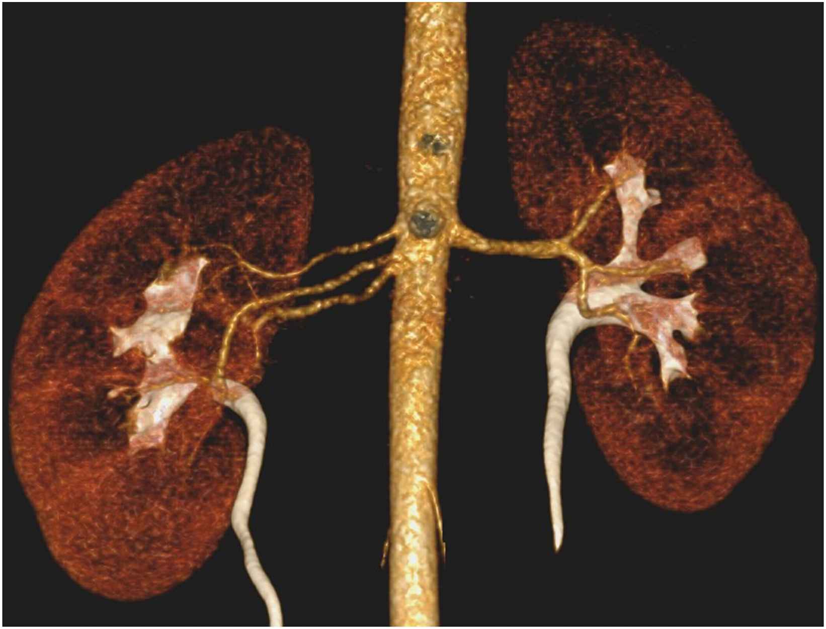
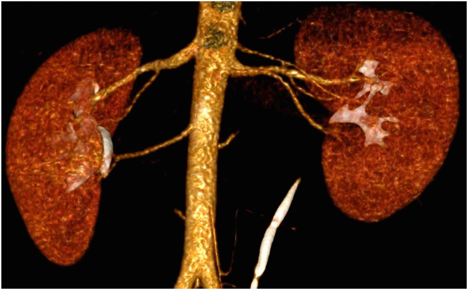
Other less common variations that we have found are the presence of multiple renal arteries (doubles or triples arteries) (Figs. 1 and 2). Some studies have reported prevalence ranging from 2 to 14%, which is the lower prevalence found in our study. Clinically this variation makes any surgical procedure involving kidney harder because the needs to do the vascular an anatomists twice, and some studies have reported more complications and grafting loss from those patients that received a kidney with anatomical variations.21
Triple RA for the right kidney.
Double RA and hiliar RA for the left kidney, and hiliar RA for the right kidney.
The prevalence of RA in our population with morphology compatible with FMD remains within the limits of what has been reported in other studies between 3 and 5%, affecting more women,22 but it was found that more patients were asymptomatic at the time of diagnosis. This is important because early detection of this disease, could probably lead to a better prognosis.23
Considering the high prevalence of these variations and the clinical importance of knowing about such variations, worldwide protocols for abdominal organ transplants has been modified to incorporate A-CT prior to the surgery (46), preventing negative outcomes related to this.
Limitations
Information is limited from one local clinic database with patients from the same hospital, however this is one of the few studies in middle and low income countries, and the only one in Colombia.
Conclusions
Variations are almost as frequent as normal anatomy in the RA; knowing their existence could help the surgeon or the interventional radiologist to avoid catastrophic complications and make a safer surgery for the patient. Additional studies are needed in different clinical settings.
Financial support
Authors declare no financial support for this research.
Ethical committee
This study was approved by our ethical committee, and does comply with the laws in Colombia.
Conflict of interests
None declared.
References
Cite this article
TY  - JOUR
AU  - Juan S. Calle Toro
AU  - Gabriel Prada
AU  - Sara Yukie Rodriguez Takeuchi
AU  - Robinson Pachecho
AU  - Gloria Baena
AU  - Ana M. Granados
PY  - 2016
DA  - 2016/02/22
TI  - Anatomic variations of the renal arteries from a local study population using 3D computed tomography angiography reconstruction images from a reference hospital in Cali, Colombia
JO  - Artery Research
SP  - 22
EP  - 26
VL  - 14
IS  - C
SN  - 1876-4401
UR  - https://doi.org/10.1016/j.artres.2016.02.004
DO  - 10.1016/j.artres.2016.02.004
ID  - CalleToro2016
ER  -