
2.6 FEASIBILITY OF AORTIC WAVE INTENSITY ANALYSIS FROM SEQUENTIALLY ACQUIRED CARDIAC MRI AND NON-INVASIVE CENTRAL BLOOD PRESSURE
- DOI
- 10.1016/j.artres.2018.10.029How to use a DOI?
- Abstract
Background: Wave intensity analysis (WIA) in the aorta offers important clinical and mechanistic insights but is difficult non-invasively. We performed WIA by combining high temporal resolution cardiovascular magnetic resonance (CMR) flow velocity and non-invasive central blood pressure (BP) waveform data.
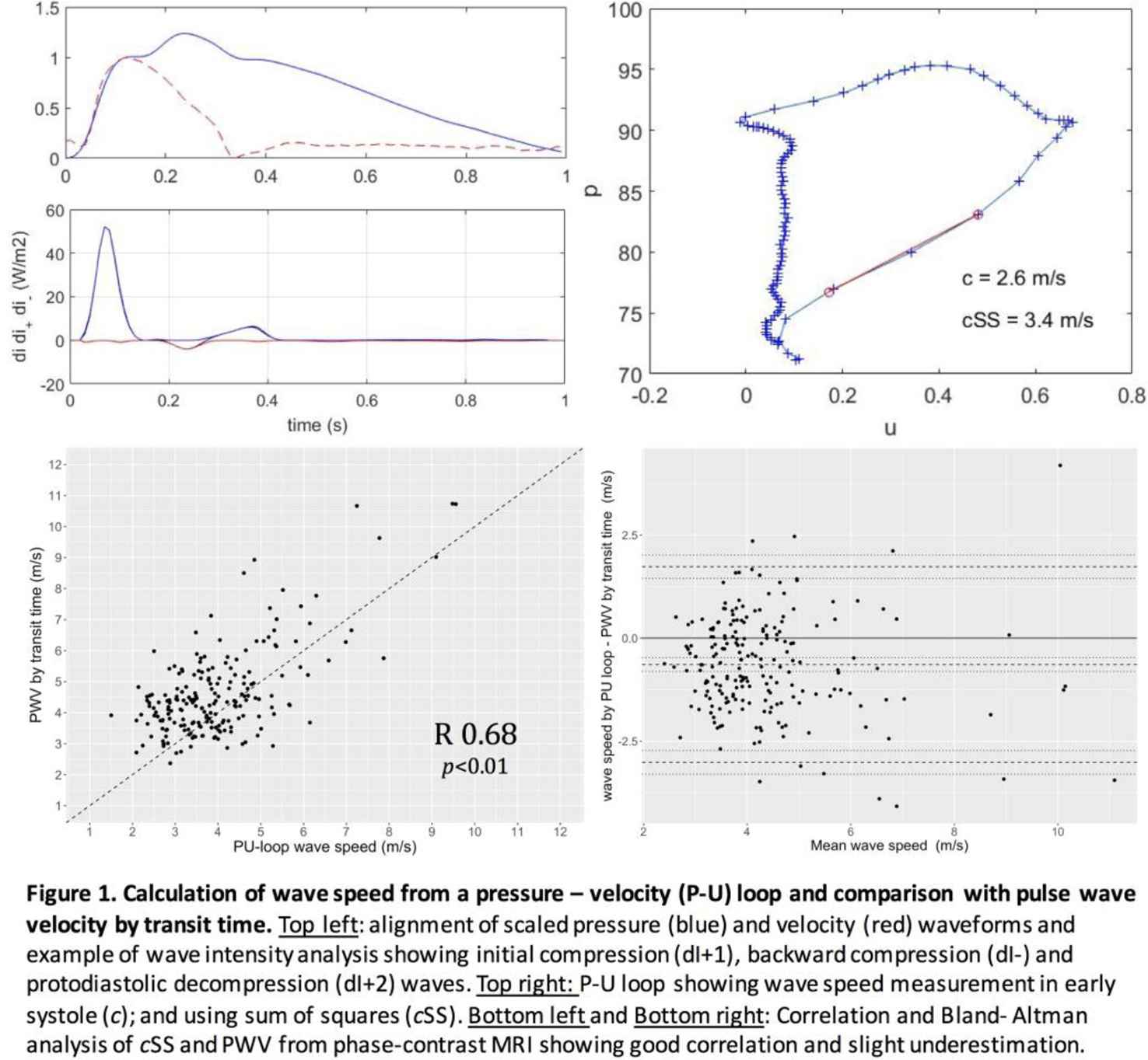
Method: 206 healthy volunteers (36 ± 11 years, 47% male) underwent sequential phase contrast CMR (Siemens Aera 1.5T, 1.97 x 1.77 mm2, ∼9 ms temporal resolution) and supra-systolic oscillometric central BP (Uscom Ltd BP+) measurement. Velocity (U) and central pressure (P) waveforms (200 Hz) were aligned using the wave foot, and local wave speed was calculated both from the P-U slope during early systole (c) and the sum of squares method (cSS) (Figure 1), and compared with CMR aortic arch pulse wave velocity (PWV) by transit time.
Results: The peak intensity of the initial compression wave (dI+1), backward compression wave (dI-) and protodiastolic decompression wave (dI + 2) were 69.5 ± 28, −6.6 ± 4.2 and 6.2 ± 2.5 W/m2 respectively. PWV correlated with c or cSS (r = 0.60, and 0.68 respectively; bias −1.3 [limits of agreement: −3.8 to 1.2 m/s], and bias −0.64 [limits of agreement: −3.0 to 1.7 m/s] respectively), Figure 1.
Conclusion: Wave intensity patterns and values are similar to those measured using invasive methods. Local wave speed showed good agreement with PWV. CMR and central blood pressure provides a novel non-invasive technique for performing wave intensity analysis and is feasible for large scale studies.
- Open Access
- This is an open access article distributed under the CC BY-NC license.
Cite this article
TY  - JOUR
AU  - Anish Bhuva
AU  - Niro Nadarajan
AU  - Andrew D’Silva
AU  - Camilla Torlasco
AU  - Redha Boubertakh
AU  - Siana Jones
AU  - Paul Scully
AU  - Rachel Bastiaenen
AU  - Guy Lloyd
AU  - Sanjay Sharma
AU  - James Moon
AU  - Kim Parker
AU  - Charlotte Manisty
AU  - Alun Hughes
PY  - 2018
DA  - 2018/12/04
TI  - 2.6 FEASIBILITY OF AORTIC WAVE INTENSITY ANALYSIS FROM SEQUENTIALLY ACQUIRED CARDIAC MRI AND NON-INVASIVE CENTRAL BLOOD PRESSURE
JO  - Artery Research
SP  - 71
EP  - 71
VL  - 24
IS  - C
SN  - 1876-4401
UR  - https://doi.org/10.1016/j.artres.2018.10.029
DO  - 10.1016/j.artres.2018.10.029
ID  - Bhuva2018
ER  -