
2.3 BIOMECHANICAL AND STRUCTURAL QUANTIFICATION OF VASCULAR DAMAGE: A UNIQUE INVESTIGATION OF STENT IMPLANTATION
- DOI
- 10.1016/j.artres.2017.10.025How to use a DOI?
- Abstract
The most challenging complication after coronary stent implantation is in-stent restenosis [1], which is mainly caused by mechanically induced injuries due to overloading. From a biomechanical point of view, the processes occurring inside the arterial tissues during stent implantation (SI) is rather unknown.
This study shows a novel approach to quantify vascular damage due to SI a multi-scale examination of coronary arteries with generated injuries using a unique experimental in vitro setup.
The setup consists of a biaxial tensile testing stage to apply physiological loads on rectangular specimens of coronary arteries and a triple-axis-unit, which allows the indentation of stent struts into arterial tissues under a specified pressure (Fig. A). In addition, the multi-scale investigation of the mechanical and structural responses of the resulting lesion, following the protocol of Sommer et al. [2], is carried out by calculating Cauchy stresses and analyzing healthy and injured specimens with second harmonic generation (Fig. B) and electron microscopy.
The results indicate that the usually wavy collagen fibers straightened, compress and align around the lesion (Fig. B). In addition, the evaluation of the material characteristics reveals a significant softening of injured tissues.
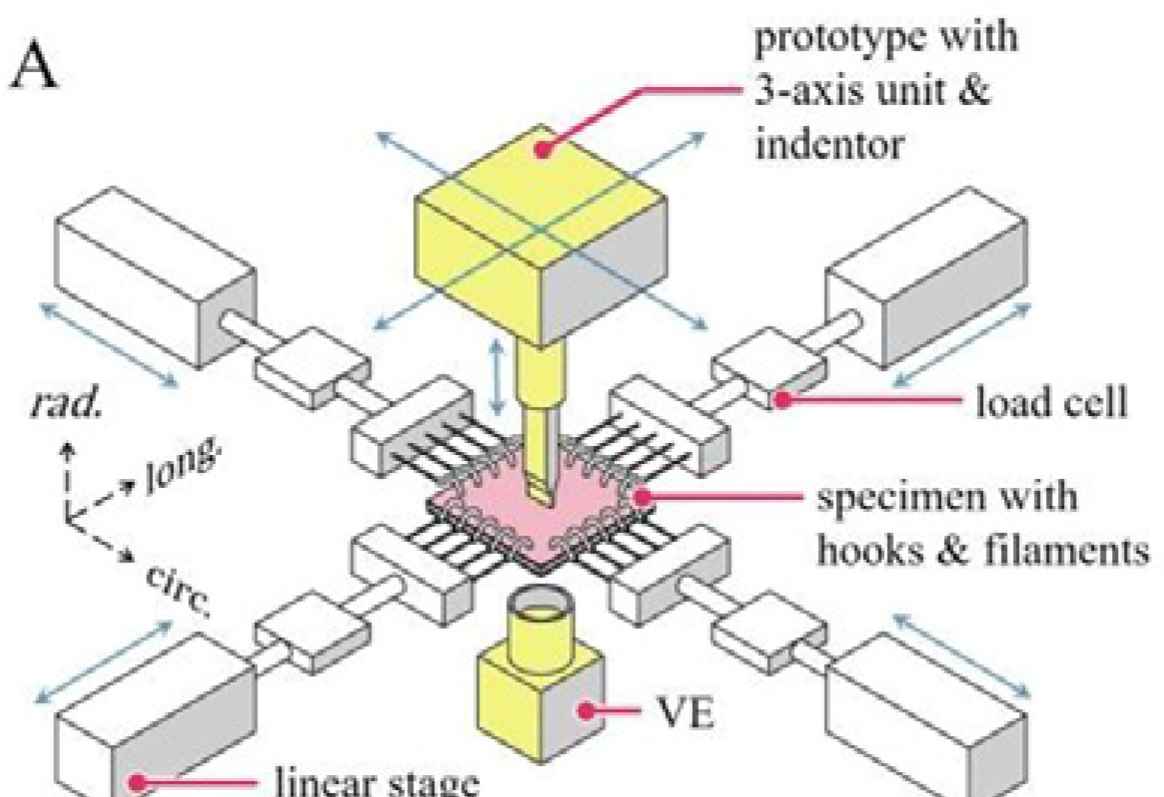 Fig. A:
Design of the experimental setup, showing a biaxial tensile testing stage (white parts) and the triple-axis-unit for indentation tests (yellow parts).
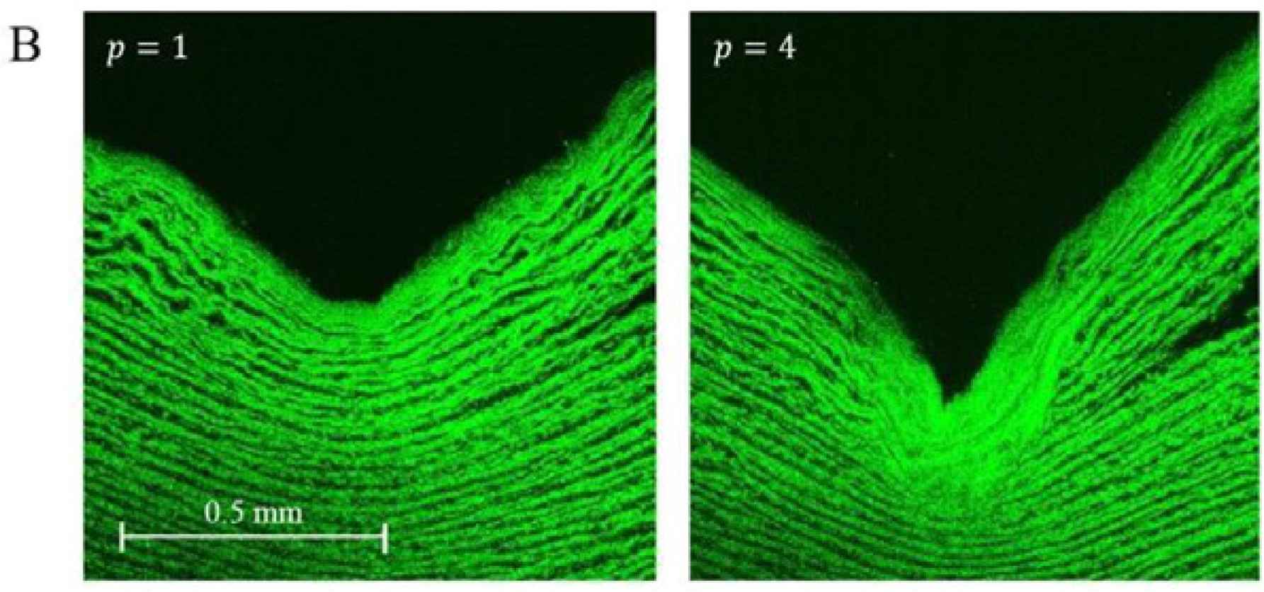 Fig. B:
Sectional view through the tissue perpendicular to the lesion. The SHG images show collagen fibers of specimens from a 6-months-old porcine descending aorta responding under different pressures (1 and 4 MPa).
- Open Access
- This is an open access article distributed under the CC BY-NC license.
Download article (PDF)
View full text (HTML)
Cite this article
TY  - JOUR
AU  - Markus A. Geith
AU  - Gerhard Sommer
AU  - Thomas Schratzenstaller
AU  - Gerhard A. Holzapfel
PY  - 2017
DA  - 2017/12/06
TI  - 2.3 BIOMECHANICAL AND STRUCTURAL QUANTIFICATION OF VASCULAR DAMAGE: A UNIQUE INVESTIGATION OF STENT IMPLANTATION
JO  - Artery Research
SP  - 50
EP  - 50
VL  - 20
IS  - C
SN  - 1876-4401
UR  - https://doi.org/10.1016/j.artres.2017.10.025
DO  - 10.1016/j.artres.2017.10.025
ID  - Geith2017
ER  -