
P80 IDENTIFICATION OF RADIAL VASCULAR WALL ABNORMALITIES BY VERY-HIGH FREQUENCY ULTRASOUND IN PATIENTS WITH FIBROMUSCULAR DYSPLASIA: THE FUCHSIA STUDY
- DOI
- 10.1016/j.artres.2017.10.096How to use a DOI?
- Abstract
Aim: This case-control study is aimed at identifying radial vascular wall abnormalities in patients with fibromuscular dysplasia (FMD).
Methods: High-frequency ultrasound scans of radial arteries were obtained by VevoMD (70 MHz probe, FUJIFILM, VisualSonics). Radial wall showed two echogenic interfaces: the 1st (lumen-media) and the 2nd (media-adventitia). Intima-media (IMT), adventitia (AT), and global thickness (IMAT) and wall cross-sectional area (WCSA) Measured. Vascular wall disarray was assessed calculating the root mean square error (RMSE) between 20 gray-level profiles crossing the two interfaces and the profile obtained averaging them, normalized for the maximum value of the corresponding mean profile (RMSE/mean).
Results: 11 treated hypertensive female FMD patients and 8 healthy controls (C) were enrolled (age 52±5 vs 45±13years, p = 0.51; BMI 26±3 vs 23±3kg/mg, p = 0.12; mean BP 97±7 vs 85±10 mmHg, p = 0.01). Radial lumen was similar; IMT (0.166±0.037 vs 0.128±0.022mm, p = 0.03), AT (0.114±0.029 vs 0.083±0.019mm, p = 0.008) and IMAT (0.281±0.042 vs 0.211±0.027mm, p = 0.003) were higher in FMD. Wall/lumen ratio was similar and WCSA increased in FMD.
The maximum values of 1st (121±43 vs 157±22, p = 0.09) and 2nd interface 109±44 vs133±18, p = 0.09) tended to be lower, whereas RMSE/mean was higher in FMD (1st 1.31±0.24 vs 0.83±0.32, p = 0.006; 2nd 1.37±0.38 vs 0.94±0.32, p = 0.03). The difference was attenuated for the 1st but not for the 2nd interface when considering age and mean BP as covariates (p = 0.054 and p = 0.016 respectively).
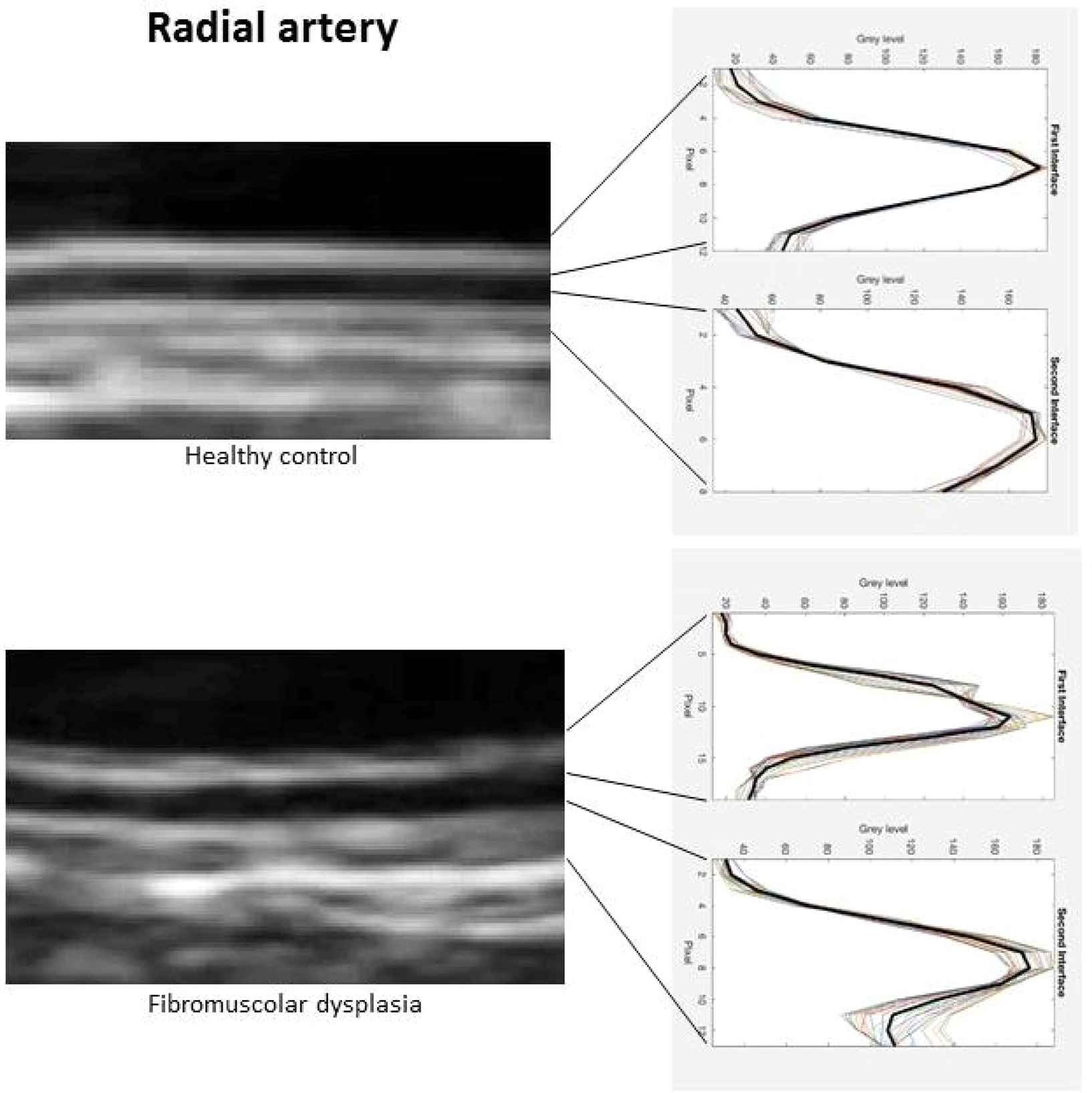
Conclusions: The radial artery wall of hypertensive FMD patients shows increased thickness and its ultrastructure is characterized of loss of echogenicity and inhomogeneity of the two echogenic layers.
- Open Access
- This is an open access article distributed under the CC BY-NC license.
Download article (PDF)
View full text (HTML)
Cite this article
TY  - JOUR
AU  - Rosa Maria Bruno
AU  - Nicole Di Lascio
AU  - Daniela Guarino
AU  - Saverio Vitali
AU  - Piercarlo Rossi
AU  - Davide Caramella
AU  - Stephane Laurent
AU  - Pierre Boutouryie
AU  - Stefano Taddei
AU  - Lorenzo Ghiadoni
AU  - Francesco Faita
PY  - 2017
DA  - 2017/12/06
TI  - P80 IDENTIFICATION OF RADIAL VASCULAR WALL ABNORMALITIES BY VERY-HIGH FREQUENCY ULTRASOUND IN PATIENTS WITH FIBROMUSCULAR DYSPLASIA: THE FUCHSIA STUDY
JO  - Artery Research
SP  - 75
EP  - 75
VL  - 20
IS  - C
SN  - 1876-4401
UR  - https://doi.org/10.1016/j.artres.2017.10.096
DO  - 10.1016/j.artres.2017.10.096
ID  - Bruno2017
ER  -