
P103 Improved Metabolic Vasoreactivity in the Brain of HM3 Patients and its Underlying Microcirculatory Mechanisms
- DOI
- 10.2991/artres.k.191224.129How to use a DOI?
- Abstract
Background: The MOMENTUM3 trial1 has revealed superiority of the novel HeartMate3 (HM3) left ventricular assist device (LVAD) compared with the HM2, with a significantly reduced occurrence of cerebrovascular accidents. Thus, cerebral autoregulation may be improved in HM3 compared with HM II patients, possibly because of altered microcirculatory haemodynamics associated with the in-built speed modulation (‘pulsatility’) of the HM3 device.
Methods: Angle-corrected Doppler ultrasound images of the middle cerebral artery (MCA) were recorded before and at the end of a 30s breathhold test in healthy controls (n = 17), heart failure (HF n = 18), HM2 (n = 10) and HM3 (n = 17) patients. Microcirculatory haemodynamics as represented by the central retinal artery (CRA) were also quantified (Controls = 33, HF = 27, HM2 = 23, HM3 = 31). Data were analysed for Time-Averaged Maximum flow velocity (TAMAX), peak flow velocity (Vmax), minimum flow velocity (Vmin), Pulsatility Index (PI) and Resistance Index (RI, Table 1).
Healthy controls (n = 33) Heart failure (n = 27) HeartMate II (n = 23) HeartMate3 (average) (n = 31) HeartMate3 (no speed modulation) (n = 31) HeartMate3 (with speed modulation) (n = 31) Middle Cerebral Artery TAMAX (cm/s) 58 ± 15 48 ± 13 45 ± 15 48 ± 19 48 ± 19 46 ± 18 Vmax (cm/s) 92 ± 22 78 ± 20 55 ± 19*# 55 ± 21*# 54 ± 21*# 55 ± 22*# Vmin (cm/s) 39 ± 12 29 ± 11 37 ± 16 39 ± 14 44 ± 16# 32 ± 13 Pulsatility index 0.88 ± 0.18 1.05 ± 0.31* 0.40 ± 0.24*# 0.34 ± 0.14*# 0.21 ± 0.12*#$ 0.56 ± 0.24*# Resistance index 0.57 ± 0.07 0.60 ± 0.11 0.29 ± 0.14*# 0.16 ± 0.09*#$ 0.17 ± 0.10*#$ 0.14 ± 0.09*#$ Central retinal artery TAMAX (cm/s) 6 ± 1 5 ± 2 6 ± 3 7 ± 3 7 ± 4 6 ± 4 Vmax (cm/s) 12 ± 3 11 ± 6 8 ± 4* 8 ± 4* 8 ± 4* 8 ± 4* Vmin (cm/s) 3 ± 1 3 ± 1 5 ± 2 5 ± 3 6 ± 3*# 4 ± 4 Pulsatility index 1.60 ± 0.45 1.42 ± 0.40 0.58 ± 0.26*# 0.49 ± 0.21*# 0.32 ± 0.17*# 0.79 ± 0.35*# Resistance index 0.75 ± 0.09 0.70 ± 0.11 0.38 ± 0.15*# 0.22 ± 0.11*#$ 0.24 ± 0.12*#$ 0.20 ± 0.13*#$ *p < 0.05 compared with healthy controls;
#p < 0.05 compared with Heart Failure;
$p < 0.05 compared with HeartMate II.
Table 1Haemodynamics in the middle cererbral artery (MCA) and the central retinal artery (CRA) between HeartMate2 and HeartMate3 patients
Results: Breathhold significantly increased TAMAX, Vmax and Vmin in all groups except HM II patients (Figure 1A). Conversely, PI decreased slightly in all groups while RI was maintained. The greater breathhold response in HM3 compared with HM2 patients was not attributable to the in-built pump-speed modulation (Figure 1B), however, HM3 had a consistently lower RI in the MCA and CRA.
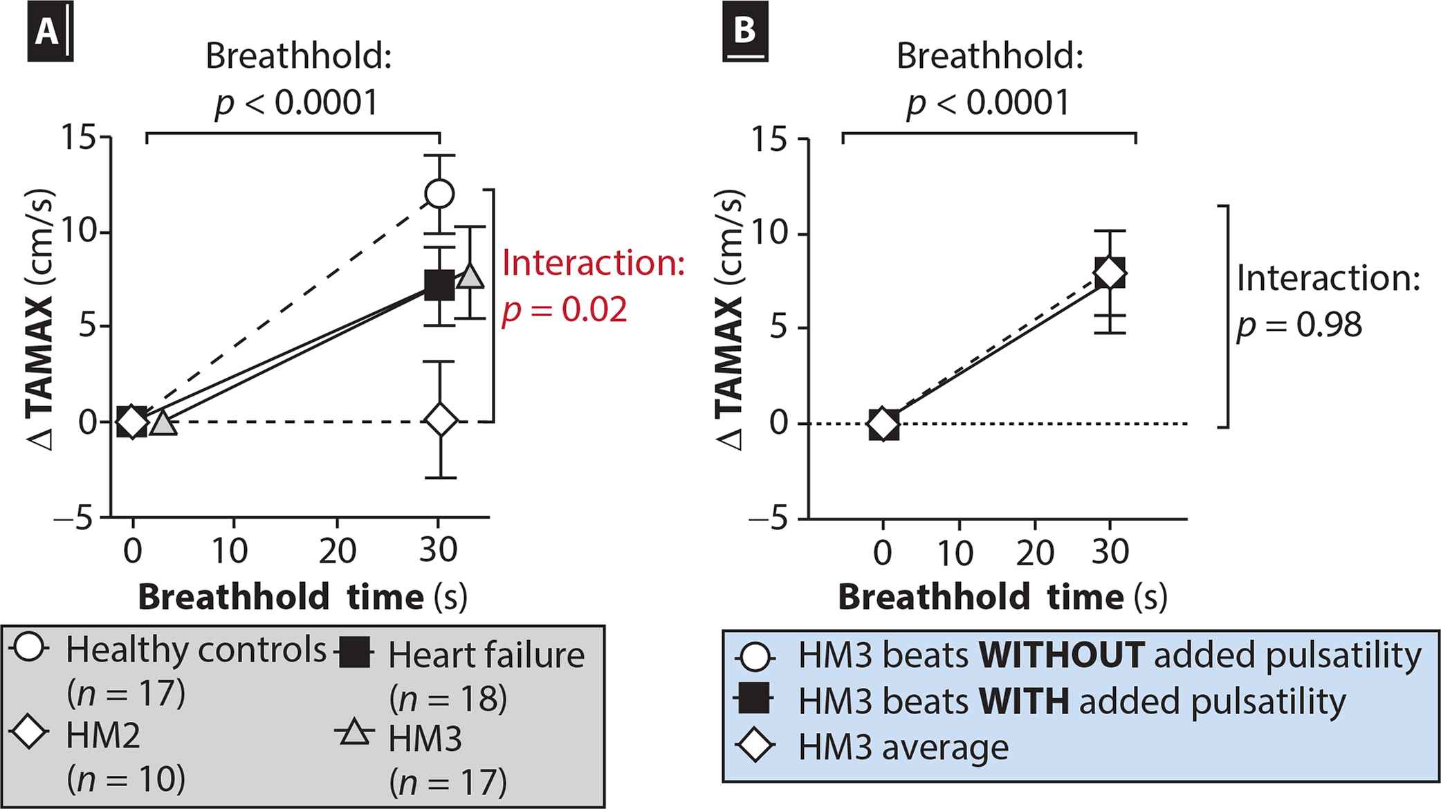 Figure 1
(A) Time-averaged maximum flow velocity in the middle cerebral artery of healthy controls and patient groups in response to a 30-s breathhold test. (B) Breakdown of the responses in HM3 patients, comparing beats with and without added pulsatility.
Conclusion: Although reduced compared with healthy controls, HF and HM3 patients have a significantly greater metabolic cerebral vasoreactivity compared with HM2 patients. The 60% greater diastolic flow velocity in the microcirculation of both LVAD groups compared to healthy controls may alter gas exchange in the microcirculation. Future studies should examine the role of altered RI in HM3 patients.
- Copyright
- © 2019 Association for Research into Arterial Structure and Physiology. Publishing services by Atlantis Press International B.V.
- Open Access
- This is an open access article distributed under the CC BY-NC 4.0 license (http://creativecommons.org/licenses/by-nc/4.0/).
Cite this article
TY  - JOUR
AU  - Eric J. Stöhr
AU  - Ruiping Ji
AU  - Koichi Akiyama
AU  - Francesco Castagna
AU  - Pinsino Alberto
AU  - John Cockcroft
AU  - Melana Yuzefpolskaya
AU  - Reshad Garan
AU  - Veli Topkara
AU  - Hiroo Takayama
AU  - Koji Takeda
AU  - Yoshifumi Naka
AU  - Paolo Colombo
AU  - Joshua Willey
AU  - Barry J. McDonnell
PY  - 2020
DA  - 2020/02/17
TI  - P103 Improved Metabolic Vasoreactivity in the Brain of HM3 Patients and its Underlying Microcirculatory Mechanisms
JO  - Artery Research
SP  - S142
EP  - S143
VL  - 25
IS  - Supplement 1
SN  - 1876-4401
UR  - https://doi.org/10.2991/artres.k.191224.129
DO  - 10.2991/artres.k.191224.129
ID  - Stöhr2020
ER  -