
Reflections Revisited, Reinterpretation Required✩
 , Lex M. van Loon1, 4, , Nico Westerhof2,
 Main findings of this short communication were presented at the Artery 2019 meeting.
- DOI
- 10.2991/artres.k.200423.001How to use a DOI?
- Keywords
- Central pressure; wave travel; pressure wave reflection; arterial stiffness; wave speed; pulse wave velocity; return time
- Copyright
- © 2020 Association for Research into Arterial Structure and Physiology. Publishing services by Atlantis Press International B.V.
- Open Access
- This is an open access article distributed under the CC BY-NC 4.0 license (http://creativecommons.org/licenses/by-nc/4.0/).
1. INTRODUCTION
In a generally accepted view, the pressure wave travels down the aorta, is reflected, and returns to the heart [1]. An increased Pulse Wave Velocity (PWV, a measure of arterial stiffness) would cause a proportional reduction in return time [2]. Additionally, in this view, the return time is longest in the central aorta and shortest in the distal aorta. However, combining data on PWV [3] and return time as a function of age [4] demonstrates that PWV has a negligible effect on return time. Moreover, the delay between the forward and reflected wave remains rather constant along the aorta [5].
2. AIMS
We aim to show why increased PWV does not reduce return time proportionally, as reported by Baksi et al. [4] and why, by the same concept, the reflected wave is not more delayed in the central aorta than distally, as presented (in the dog) by Tyberg et al. [5].
3. MATERIALS AND METHODS
We used two models of the systemic arterial system based on oscillatory flow theory [6]: a single uniform tube model (“single tube”; with a Windkessel load) representing the aorta, and a distributed (1D) model of the entire arterial tree (“whole system”). We derived forward and reflected waves in the aorta following the standard method [7], to evaluate the return time of the reflected wave with increasing PWV. Also, forward and reflected waves along the aorta of both models were calculated. Input impedance and reflection coefficient along the model-aorta were derived. Detailed model descriptions are given in Westerhof and Westerhof [8].
4. RESULTS
Central pressures with forward and reflected waves in the uniform tube and in the whole system were calculated for increasing PWVs (Figure 1). In the uniform tube, the central arrival time of the reflected wave is inversely related to the PWV, while in the whole system there is very little effect of increasing PWV.
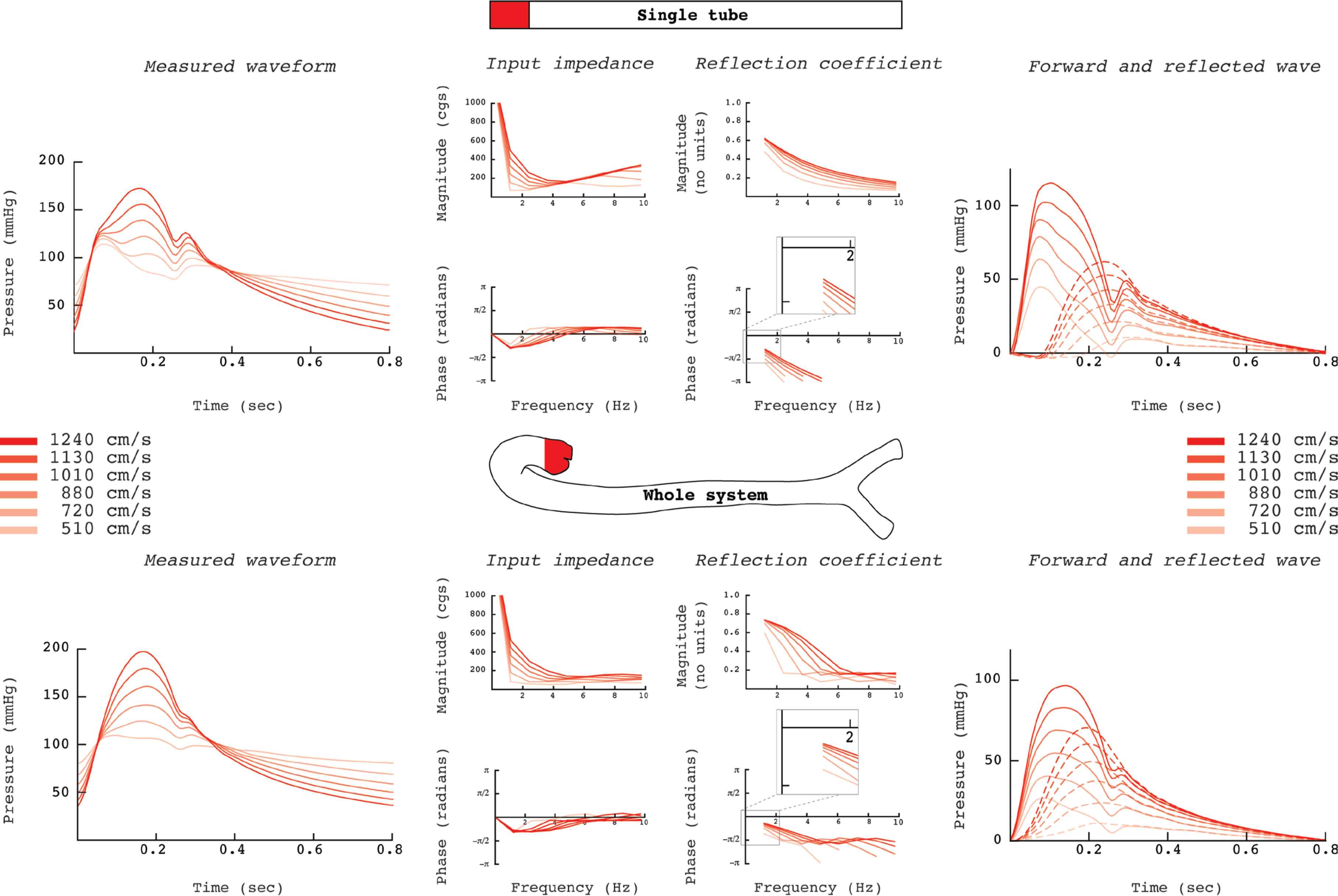
Central pressures with forward and reflected wave in the uniform tube and in the whole system for increasing pulse wave velocities (cgs indicates units in the centimetre-gram-second system). In the single tube model, the reflected wave arrives considerably earlier at the central aorta for increased pulse wave velocities, while in the whole system, reflected wave arrives sooner and the effect of increasing Pulse Wave Velocity (PWV) is minimal. This follows from the behaviour of the input impedance, which, together with the characteristic impedance of the supplying vessel, determines the reflection coefficient. In the uniform tube, increasing PWV has more impact on the phase of the input impedance and thus causes substantial delay of the reflected wave for low PWVs, which decreases with increasing PWV. In the whole system this effect is less pronounced: the reflected waves start increasing with a comparable delay. Thus, return of the reflected wave hardly changes as was shown by Baksi et al. [4]. The pulse wave return time was “calculated from the time of the shoulder on the pressure waveform” [4]. Although this “shoulder point” is most likely determined by the return of the reflected wave, it should be noted that the assessment of the return time of the reflected wave from measured pressure is not straightforward, since neither “inflection point” nor “shoulder point” [9] are related to the reflected wave in a simple way [10]. In the whole system, the reflection coefficient increases with PWV in the lower frequency range. With it, the relative contribution of the reflected wave increases, and this in turn increases the pressure augmentation. Thus, perhaps surprisingly, the pressure augmentation is more related to PWV than is the return time.
Pressures along the uniform tube and the aorta of the whole system and the corresponding forward and reflected waves were calculated (Figure 2) at a PWV of 720 cm/s. The uniform tube model predicts a reflected wave that travels proximally from the tube end, a backward running wave. The 1D model shows that the reflected wave is at all locations delayed with respect to forward wave.
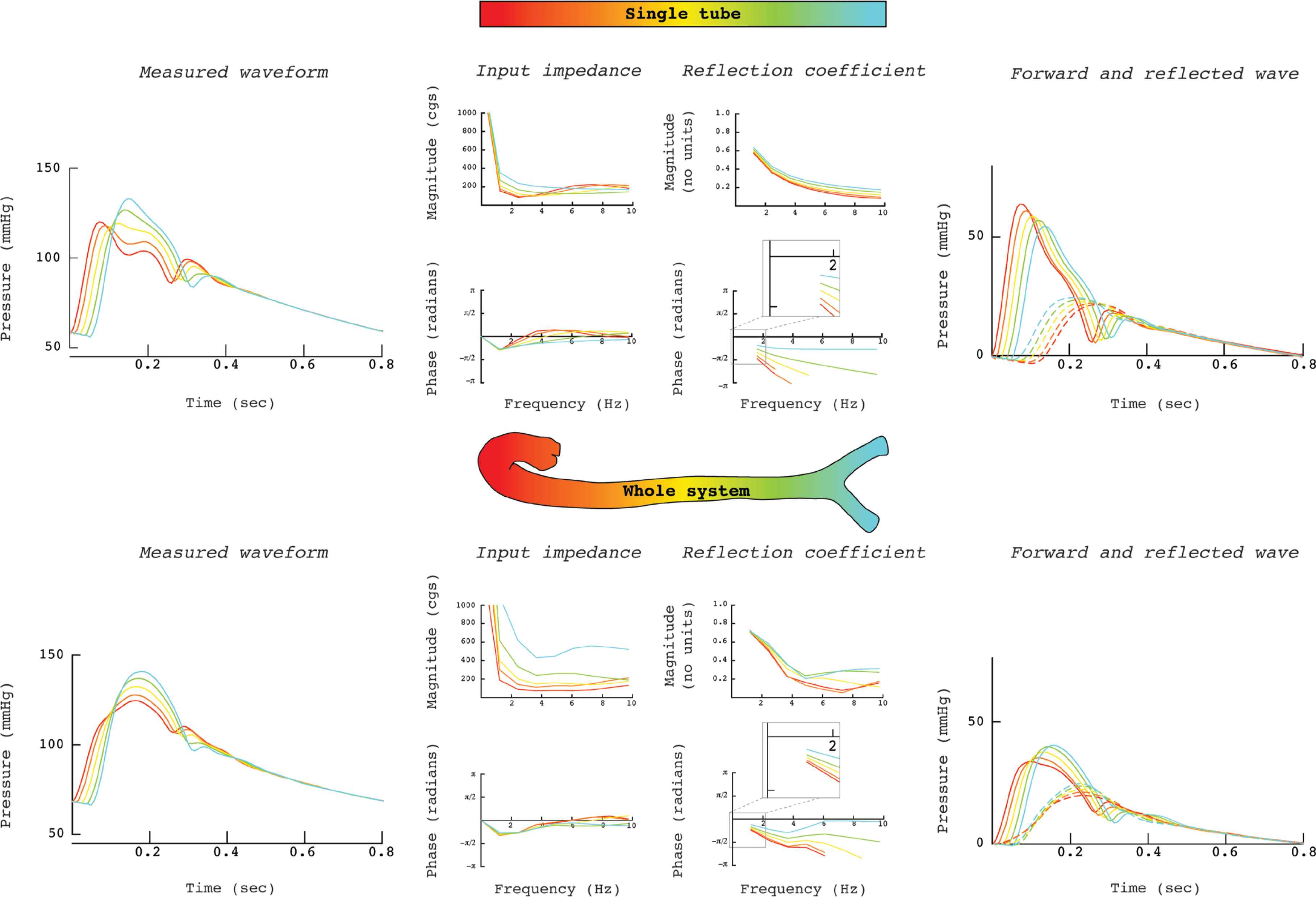
Pressures along the uniform tube and the aorta of the whole system and the corresponding forward and reflected waves; Pulse Wave Velocity (PWV) is kept constant (cgs indicates units in the centimetre-gram-second system). In the tube model, the delay between the forward and the reflected wave is large, while the delay is small close to the distal reflection site. In the whole system, in the central aorta the reflected wave arrives sooner, and the delay is comparable at the more distal sites. This can be understood by features of the input impedance, which, together with the characteristic impedance of the supplying vessel, determine the reflection coefficient. In the uniform tube, the differences in the phase of the reflection coefficients are considerable for the separate sites. These result in large delay of the reflected wave for the proximal sites, and less delay for the distal sites. In the whole system, this effect is less prominent since the difference in the phase is much less.
5. CONCLUSION
In the human aorta, the return time of the reflected wave is not inversely proportional to PWV [4]. Also, reflected waves have a relatively constant delay with respect to the forward wave, appearing to run downstream rather than to the heart [5]. These findings can be explained with the 1D model but not with the single uniform tube model with a distal reflection site. The multiple reflection sites in the real system cause reflections and re-reflections, together leading to a compound reflected wave, which does not exist in the uniform tube model.
The delay between the forward and reflected wave in the proximal aorta is determined by the phase of the reflection coefficient, the low frequencies determining the gross description of the pressure [8]. When inaccurately describing the arterial system as uniform tube, the phase indeed shows large dependency on PWV. However, in the whole system, phase is much less dependent on PWV. Consequently, the return time of the reflected wave is not inversely proportional to PWV.
At any location in the arterial system, the characteristic impedance (Zc) of the supplying vessel and the input impedance, Zin, of the downstream system determine the reflection coefficient. Since at all locations the loading input impedance is made up of a system of many arteries with multiple reflection sites, the normalized input impedances (Zin/Zc) show a comparable spectrum [8]. Thus, the phase does not systematically increase or decrease with distance. This implies that the time difference between reflected and forward wave is not increasing towards the heart as predicted by the single-tube model.
Therefore, we advise that a single uniform tube with a distal reflection site should be abandoned as conceptual model. It erroneously suggests an earlier return time with increasing PWV (older age) and it misleadingly implies that the delay between the forward and the reflected wave is larger for more proximal sites. A frequency domain interpretation is required, where the phase of the reflection coefficient explains the findings.
CONFLICTS OF INTEREST
The authors declare they have no conflicts of interest.
AUTHORS’ CONTRIBUTION
BEW and NW conceived the study. BEW ran the simulations. BEW, NW and LMvL wrote the manuscript. All authors reviewed the final manuscript.
Footnotes
REFERENCES
Cite this article
TY  - JOUR
AU  - Berend E. Westerhof
AU  - Lex M. van Loon
AU  - Nico Westerhof
PY  - 2020
DA  - 2020/04/28
TI  - Reflections Revisited, Reinterpretation Required✩
JO  - Artery Research
SP  - 117
EP  - 120
VL  - 26
IS  - 2
SN  - 1876-4401
UR  - https://doi.org/10.2991/artres.k.200423.001
DO  - 10.2991/artres.k.200423.001
ID  - Westerhof2020
ER  -