
Neonatal Hemochromatosis: Treatment with Exchange Transfusion and Intravenous Immunoglobulin
 , Abdulrahman Al Zahrani1, Mohamed Abdoun1, , Syeda Naqvi1, Georges E. Nawfal2,
 - DOI
- 10.2991/dsahmj.k.210715.001How to use a DOI?
- Keywords
- Hemochromatosis; liver failure; gestational alloimmune liver disease; immunoglobulin; exchange transfusion
- Abstract
Neonatal Hemochromatosis (NH) is a rare phenotype of severe fetal/neonatal liver injury that is accompanied by extrahepatic siderosis. Current clinical evidence shows that NH is not a disease per se, but is the consequence of fetal liver injury. Gestational alloimmune liver disease is the cause of nearly all cases of NH. Affected babies may die in utero, or present postnatally with severe acute liver failure or decompensated congenital liver cirrhosis. Diagnosis depends on the demonstration of extrahepatic siderosis by Magnetic Resonance Imaging (MRI), buccal biopsy, or detection of complement C5b–9 complex on hepatocytes from liver biopsy. Prognosis is generally bad without treatment. Treatment with iron chelators and antioxidants is not helpful. The more recent treatment approach of exchange transfusion and Intravenous Immunoglobulin (IVIG) has shown favorable outcomes. In this report, we describe a case of NH that presented with liver cell failure and high serum ferritin. Diagnosis was confirmed by MRI by demonstration of siderosis in the liver and pancreas while sparing the spleen. The infant was successfully treated with a combination of exchange transfusion and IVIG, and discharged at age 30 days in good condition.
- Copyright
- © 2021 Dr. Sulaiman Al Habib Medical Group. Publishing services by Atlantis Press International B.V.
- Open Access
- This is an open access article distributed under the CC BY-NC 4.0 license (http://creativecommons.org/licenses/by-nc/4.0/).
1. INTRODUCTION
Neonatal Hemochromatosis (NH) is a rare condition that was initially described in 1957 by Cottier and Vogt [1]. Unlike hereditary hemochromatosis, NH is a secondary condition rather than a primary iron storage disease [1–3]. Liver injury in NH causes poor regulation of maternofetal iron flux that eventually results in iron overload. Current evidence suggests an underlying alloantibody-mediated liver injury in almost all cases of NH (Gestational Alloimmune Liver Disease; GALD), with secondary altered iron metabolism (siderosis) [4,5].
Prognosis is poor and treatment methods are limited. Previously, an approach that included a combination of antioxidants and iron chelator was disappointing with a reported success rates of only 10–20% [6,7]. A recent approach based on the alloimmune nature of the disease was introduced in 2009, which includes Exchange Transfusion (ET) and Intravenous Immunoglobulin (IVIG). Results from case reports and cohort studies are promising [8–10]. Liver transplantation may be needed in refractory cases [11]. We report a case of NH that was successfully treated with ET and IVIG.
2. CASE REPORT
We report a 2.5 kg female baby delivered by cesarean section at 36 weeks of gestation from consanguineous parents. APGAR scores were 8 and 9 by 1 and 5 minutes, respectively. She was admitted to the Neonatal Intensive Care Unit (NICU) on high-flow nasal cannula due to tachypnea which improved over 24 h. Her mother was gravida 4, and her first pregnancy ended in early miscarriage. A 4-year-old boy was the outcome of the second pregnancy. The third pregnancy gave rise to a full-term female baby who was admitted to the NICU with signs of acute liver failure that was suspected to be due to tyrosinemia but she died at age 40 days before confirming the diagnosis.
In our case, the initial investigations revealed signs of liver cell failure: total serum bilirubin 252 μmol/L, direct bilirubin 95 μmol/L, Aspartate Transaminase (AST) 118 IU/L, Alanine Transaminase (ALT) 36 IU/L, albumin 17 g/L, hypoglycemia, and unrecordable Partial Thromboplastin Time (PTT), Prothrombin Time (PT) and International Normalization Ratio (INR). Abdominal ultrasound showed mild ascites and there was peripheral limb edema. Initial platelets count was 37,000. Serum α-fetoprotein was >20,000 ng/mL (normal range: 10–20 ng/mL). The infant was kept on intravenous fluids, nil by mouth, and started on vitamin K supplementation. Although there was no evident bleeding, the deranged coagulation profile and thrombocytopenia were prophylactically corrected with Fresh Frozen Plasma (FFP) daily for 6 days and platelet transfusion daily for 8 days. At the same time, a workup to uncover the underlying cause was initiated, stressing initially on metabolic disorders because of the consanguineous marriage and history of a previous sibling with suspected tyrosinemia. The patient was started on ampicillin and cefotaxime until sepsis was ruled out. Markers for infection, including blood culture, white blood cell count, and C-reactive protein, were benign.
Both serum ammonia and lactate were mildly elevated (126 μmol/L and 2.6 mmol/L, respectively). Metabolic screening was unremarkable except for elevated phenylalanine level (161 μmol/L, normal range: 18–131 μmol/L). Succinylacetone was absent in urine both before and after starting feeding. Similarly, metabolic screening for galactosemia was unremarkable. Serum ferritin was high (3461 ng/mL, normal range 25–200 ng/mL). All these findings directed us to the possibility of NH.
Abdominal Magnetic Resonance Imaging (MRI) was compatible with the diagnosis of NH. It showed clear siderosis that affected the liver and pancreas while sparing the spleen (Figure 1).
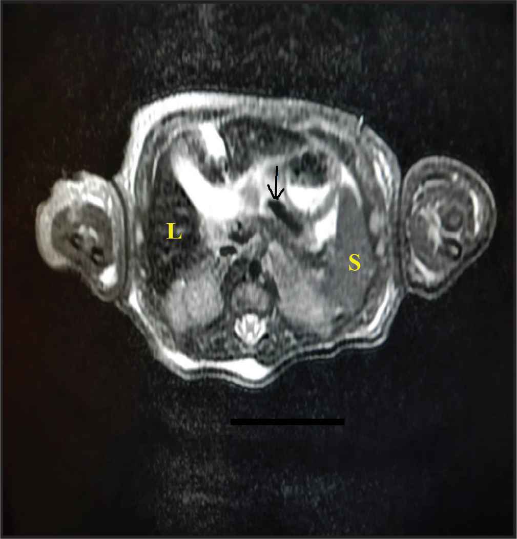
Abdominal magnetic resonance imaging. There was a dark T2 signal on the liver (L) and to a lesser extent, pancreas (arrow), compared to the skeletal muscles. The spleen (S) showed normal signal intensity. This was suggestive of neonatal hemochromatosis.
Double-volume ET was conducted at age 5 days followed by IVIG, 1 g/kg, daily for 3 doses. Platelet transfusion and FFP were discontinued by day 8 and gradual feeding was started on day 9. The infant showed gradual improvement over the 30 days she spent in NICU.
By the time of discharge, her liver profile showed serum albumin 27 g/L, AST 59 IU/L, ALT 26 IU/L, PTT 37 s, PT 12 s, INR 1.1, direct bilirubin decreased to 77 μmol/L, and platelet count reached 95,000. Her ferritin dropped to 862 ng/mL. She was discharged at age 30 days on regular milk formula and in a good general condition for follow-up in the outpatient clinic. Her family was instructed about the importance of antenatal IVIG in future pregnancies.
3. DISCUSSION
Neonatal hemochromatosis is a rare, severe liver injury with onset in the perinatal period and is characterized by deposition of stainable iron in both hepatic and extrahepatic tissues [12]. Initially, NH was considered as a form of hereditary hemochromatosis, which is a common autosomal recessive disorder with excessive iron deposition in the liver as well as in extrahepatic tissues that is usually present above 40 years of age [1,2]. A genetic defect was also suspected due to the recurrence in subsequent offspring but investigation has so far failed to prove this theory [5]. Recent evidence supports that most cases of NH are due to an alloimmune disease and the term NH is currently replaced by GALD-NH. Similar to other maternofetal alloimmune diseases, GALD is mediated by maternal Immunoglobulin G (IgG) that crosses the placenta around the 12th week of gestation after being sensitized to fetal hepatocyte antigens [1,3,13,14].
Extrahepatic siderosis in NH is most frequently seen in the pancreas, myocardium, thyroid gland, and the mucosa of minor salivary glands of the oronasopharynx. Parathyroid glands, pituitary gland, endocrine pancreas and renal tubules are less frequently affected. The reticuloendothelial system is relatively spared [15]. In fact, the siderosis seen in NH is secondary to abnormal iron handling by the injured fetal liver [13].
It is still unclear how mothers are exposed to fetal hepatic antigen. Once the IgG crosses to the fetus, it binds to a hepatocyte antigen and activates terminal complement reaction with the formation of the membrane attack complex. The C5b–9 complex that is generated during this process can be demonstrated by immunohistochemical staining in nearly all hepatocytes of infants with GALD-NH [3]. In <2% of cases, NH is due to non-GALD causes that include metabolic, syndromic, and infectious causes [16]. Affected liver shows marked or even total loss of hepatocytes [4,15]. Surviving hepatocytes show coarsely granular siderosis. Severe panlobular fibrosis and cirrhosis are common features. Immunohistochemical staining for C5b–9 helps to detect the membrane attack complex in liver specimens [17].
Our case presented with both clinical and laboratory signs of liver cell failure immediately after birth. NH is one of the most common causes of neonatal acute liver failure. It can present any time from 16 weeks’ gestation to 3 months after delivery but most cases present in first few postnatal hours [18,19].
In our case, there was a family history of similar liver cell failure in a previous sibling. GALD is reported to occur in the first pregnancy with a recurrence rate more than 90% in subsequent pregnancies [20].
As in our case, affected infants may be small for gestational age, premature and edematous. They often have jaundice, hypoalbuminemia, hypoglycemia, and abnormal coagulation profile. Disease severity varies among affected infants [21,22]. Both conjugated and unconjugated bilirubin is elevated. Direct bilirubin may be only modestly elevated. Aminotransferases rarely exceed 100 IU/L. α-Fetoprotein levels are high (100,000–600,000 ng/mL) [4,6]. Serum ferritin levels are also high (>800 ng/mL), and it is a sensitive rather than a specific indicator of NH [15].
The clinical and laboratory signs of liver cell failure as well as the high ferritin level in our case were clues to the diagnosis of liver cell damage. The family history of one sibling that had died in early neonatal life with a possible diagnosis of tyrosinemia made us consider the possibility of a metabolic disorder in the present case. A workup to uncover the underlying cause of liver cell failure was started, including screening for metabolic disorders and tests for sepsis and congenital infections. Metabolic screening was done to exclude metabolic causes of liver cell failure such as galactosemia, tyrosinemia, and mitochondrial diseases. The screening was unremarkable except for mildly elevated phenylalanine without positive succinylacetone in urine. Infants with NH may have elevated phenylalanine and tyrosine levels, but they do not have succinylacetone in the urine. Lactate should not be markedly elevated [17].
Gestational alloimmune liver disease-neonatal hemochromatosis was also considered in our case. Clues for the diagnosis of GALD-NH were the clinical and laboratory features of liver cell failure, family history of a similar condition, high ferritin level, and abdominal MRI that showed evidence of siderosis in liver and pancreas and not the reticuloendothelial system (spleen). Tissue biopsy was not done due to the markedly deranged coagulation profile. Liver biopsy was not done because of the positive MRI findings, risk of bleeding, and the lack of facilities for immunohistochemical study.
Gestational alloimmune liver disease-neonatal hemochromatosis should be suspected in neonates presenting with acute liver failure or congenital cirrhosis. It should also be considered in cases of Unexplained Intrauterine Fetal Death (IUFD), stillbirth, or early neonatal death. The clinical and laboratory evidence of liver disease combined with evidence of extrahepatic hemosiderosis were traditionally the clues for diagnosis of NH [17]. However, as well as evidence that supports the alloimmune nature of NH, the characteristic immunohistological features also help in the diagnosis [23].
Siderosis in the liver alone is not diagnostic as the normal newborn liver can contain quantities of iron that are stainable. In addition, pathological hepatic siderosis can be seen in several neonatal liver diseases [12]. Extrahepatic siderosis can be confirmed by iron staining studies on tissue biopsies, preferably from oral mucosa and submucosal salivary glands [24].
T2-weighted MRI can also be used to document siderosis because affected tissues have different magnetic susceptibility than normal tissue has, particularly in the liver and pancreas, myocardium and thyroid gland. In fact, MRI is the most helpful noninvasive study in the diagnosis of NH. It detects abnormal hepatic and pancreatic iron levels with absent siderosis in the spleen [25,26]. It is the quickest and least invasive method for demonstration of extrahepatic siderosis. A moderate-to-severe degree of pancreatic siderosis tends to be associated with NH [27].
Some experts have reported that each adequate oral biopsy and T2-weighted MR image has a sensitivity of 60% if used separately and 80% if used together in demonstrating siderosis in NH. A suggested approach for diagnosis is to start with oral biopsy or MRI and only if that test is negative should the other be performed. If extrahepatic siderosis cannot be demonstrated, liver biopsy for C5b–9 staining can be considered [17].
Prognosis without treatment is poor. Our case received supportive treatment to correct the abnormal coagulation profile and thrombocytopenia. Supportive care includes transfusion of FFP, platelets, cryoprecipitate, and packed red blood cells [7].
The current recognition of the alloimmune nature of NH has led to a new treatment approach that involves a combination of double-volume ET followed immediately by administration of high-dose IVIG (1 g/kg). ET helps to remove reactive antibody while IVIG blocks antibody-induced complement activation [28].
After establishing the diagnosis of GALD-NH, we started a specific treatment with combination of ET and three doses of IVIG. Studies published over the past few years have shown marked cure rates with this regimen. Heissat et al. [29] presented a primary diagnostic work-up that included lip biopsy and/or MRI. They suggested ET and IVIG before liver biopsy if a specific viral, metabolic, or hypoxemic cause of neonatal acute liver failure cannot be identified. Others recommended that any infant with liver failure should be given one dose of IVIG while NH is being considered. If NH is proven by MRI and/or lip biopsy, and the infant has not improved, an exchange transfusion should be performed, followed by administration of a second dose of IVIG [8,17,30]. There is no standard regimen for frequency of IVIG treatment of GALD-NH, and it depends on the degree of improvement in clinical and laboratory parameters. Regimens with one to three doses have been reported [8,10,17,29,31–33]. ET alone has been reported to be effective in some cases of GALD [9].
Previously and before knowing the alloimmune nature of NH, based on the concept that NH was secondary to oxidative injury caused by hemosiderosis, a cocktail of antioxidants and an iron chelator was used. The results of this approach were disappointing with success rates as low as 10–20% [30]. This is why we did not consider antioxidant and iron-chelating therapy in our case.
Liver transplantation, although the only curative approach in the past, was challenging because of the nature of associated morbidity including prematurity, low birthweight, and multisystem failure. Survival rate was only about 35%. It is also difficult in this age group due to lack of appropriate donors and the increased risk of vascular and infectious complications [34].
With medical treatment, recovery of the injured liver may take 4–6 weeks [8]. Most infants can go home in 1–4 months. However, it may take 2–4 years for the liver to fully recover. Long-term outcome after medical treatment is still a matter of observation [17]. Although our case was sufficiently recovered to go home by age 30 days, we informed her parents that full recovery may need several months and long-term follow-up in the clinic would be mandatory.
Our case was the second affected baby for the family; the first died early in the neonatal period and presented with signs of acute liver failure that was thought to be due to an inborn error of metabolism, mostly likely tyrosinemia. Recurrence rate of GALD-NH in subsequent pregnancies is >90%. IVIG for pregnant women with a previous pregnancy that resulted in an infant with GALD (IVIG, 1 g/kg/week beginning in week 14 of the pregnancy until the end of gestation) significantly reduces the risk of recurrence and fetal loss [20,35]. We counseled the parents of our case about the benefits of antenatal IVIG in preventing NH in future pregnancies.
4. CONCLUSION
Neonatal hemochromatosis is the most common cause of neonatal acute liver failure. NH is almost always due to GALD. It should be suspected in all neonates who present with clinical and laboratory evidence of acute liver injury, as well as in all cases of unexplained stillbirth, IUFD, or early postnatal death. Diagnosis is confirmed by demonstrating extrahepatic siderosis by MRI or tissue biopsy. Immunohistochemical studies, if available, are of great diagnostic value. A combination of IVIG and ET is currently the treatment of choice. NH has a high recurrence in subsequent pregnancies, and antenatal IVIG treatment can greatly reduce this risk.
CONFLICTS OF INTEREST
The authors declare they have no conflicts of interest.
AUTHORS’ CONTRIBUTION
EMH collected the data and prepared the manuscript. SN and MA helped with data collection. AAZ supervised the work. All the authors review the manuscript and approve the final draft.
ACKNOWLEDGMENTS
We would like to express our gratefulness to the parents of our baby who provided us with all the details of the previous sibling, and were willing to publish the data about their babies. Special thanks to all NICU staff and Dr Georges who effectively shared in the diagnosis and management of this baby.
ABBREVIATIONS
- ALT,
alanine transaminase;
- AST,
aspartate transaminase;
- ET,
exchange transfusion;
- GALD,
gestational alloimmune liver disease;
- INR,
international normalization ratio;
- NH,
neonatal hemochromatosis;
- PT,
prothrombin time;
- PTT,
partial thromboplastin time.
REFERENCES
Cite this article
TY  - JOUR
AU  - Ehab Mohamed Hantash
AU  - Abdulrahman Al Zahrani
AU  - Mohamed Abdoun
AU  - Syeda Naqvi
AU  - Georges E. Nawfal
PY  - 2021
DA  - 2021/07/27
TI  - Neonatal Hemochromatosis: Treatment with Exchange Transfusion and Intravenous Immunoglobulin
JO  - Dr. Sulaiman Al Habib Medical Journal
SP  - 99
EP  - 103
VL  - 3
IS  - 3
SN  - 2590-3349
UR  - https://doi.org/10.2991/dsahmj.k.210715.001
DO  - 10.2991/dsahmj.k.210715.001
ID  - Hantash2021
ER  -