
Paraplegia Due to Stent Fracture, after Thoracic Endovascular Aortic Repair for Blunt Thoracic Aortic Injury: A Case Report and Review of Literature
 , Ahmed Sameer Mahmud Nadeem4, , Mutaz I.K. Fakhry Al-Khateeb2, Adel Aswad3
- DOI
- 10.2991/dsahmj.k.210811.001How to use a DOI?
- Keywords
- Aorta; dissection thoracic; blunt thoracic injury; endovascular treatment; stent fracture
- Abstract
Thoracic Endovascular Aortic Repair (TEVAR) became the standard treatment for patients with Blunt Thoracic Aortic Injury (BTAI) because it is a minimally invasive intervention, with proven lower morbidity and mortality compared with open surgery, especially in multiple trauma patients. However, there is still some debate about the grade of BTAI which requires intervention, the durability of the stent, and its future complications, as well as about the age limit for the younger age group to be treated by TEVAR. We report a case of a 19-year-old male who presented to the emergency department complaining of lower chest and upper abdominal pain radiating to his back with gradual loss of motor ability at his lower limbs for the previous 2 days. These symptoms started immediately after heavy lifting and a jump from a scaffold at the patient’s workplace. The patient had a history of BTAI, associated with other injuries, treated successfully with TEVAR, and discharged from hospital 13 months ago, after an uneventful post-intervention period. He was not committed to the follow-up schedule and continued his strenuous job after being discharged. The patient was admitted, and computerized tomography angiography showed distal aortic stent fracture with thrombosis occluding the lower thoracic aorta, for which the patient was treated with another stent graft. This is the first case report of stent fracture after TEVAR in a young patient provoked by strenuous work, demonstrating an early stent fracture, which usually occurs as a late complication of TEVAR.
- Copyright
- © 2021 Dr. Sulaiman Al Habib Medical Group. Publishing services by Atlantis Press International B.V.
- Open Access
- This is an open access article distributed under the CC BY-NC 4.0 license (http://creativecommons.org/licenses/by-nc/4.0/).
1. INTRODUCTION
Blunt Thoracic Aortic Injury (BTAI) is a rare injury, reported to be about 0.3% of overall trauma admissions [1], but it is life-threatening and the second leading cause of death in all patients with nonpenetrating trauma [2,3]. The incidence of BTAI is estimated to be between 1.5% and 2% in patients who sustain blunt thoracic trauma [4,5]. The principal mechanism of the injury is usually a rapid deceleration of high-energy blunt trauma, either from a road traffic accident (in more than 70% of cases) or falls from a significant height and other causes [6–8]. Males are reported to be more subjected to BTAI [9], and it is more prevalent in obese individuals [8].
Up to 80% of BTAI patients die prior to reaching the hospital, whereas in the remaining survivors, in-hospital mortality is as high as 46% [10,11]. Because of the high mortality of BTAI, which accounts for up to one-third of reported deaths within the first 24 h, early and prompt diagnosis is very important especially with the polytrauma patient’s presentation, such as cardiac trauma, fractures (sternum, ribs, clavicle, and/or scapula), flail chest, and others [3,12]. Therefore, the diagnosis of BTAI is essentially based on the history and mechanism of trauma, clinical suspension, and radiological/imaging findings.
Clinical findings on physical examination have low sensitivity and specificity for this type of injury, although chest wall contusion and abrasions may increase suspicion [12,13]. Chest X-ray indicates the diagnosis and BTAI is confirmed using Computerized Tomography Angiography (CTA), and in rare cases Intravascular Ultrasound is needed when we have equivocal findings or for the sizing of the aorta diameter [14,15]. In an unstable patient who cannot be transferred to the radiology department for CTA and who requires resuscitation, Transesophageal Echocardiography can be used to clarify the injury [3].
Treatment of BTAI is guided by the classification of the injury [16] using the Society for Vascular Surgery criteria [17]. Apart from the conservative treatment in grade 1 injury, the treatment of BTAI in the past two decades has shifted toward Thoracic Endovascular Aortic Repair (TEVAR), because of its lower morbidity and mortality; however, the long-term outcome is still unclear and warrants further follow-up. Open surgery and combined endovascular with open surgery still have a role in the treatment of BTAI [18].
Acute aortic device-related complications (endoleak, endograft infection, false lumen expansion, stent-graft collapse, migration, deformity, fracture, and distal pseudoaneurysm) have been reported extensively [19,20]; however, there is paucity in reports of late complications, especially in young adults and after BTAI [21,22].
In this case report, who focus on a young patient who presented with paraplegia because of aortic stent fracture causing thrombosis 13 months after undergoing TEVAR for BTAI and the possible impact of strenuous type of work as a cause of the fracture.
2. CASE REPORT
A 19-year-old male patient presented to the emergency department complaining of 2 days of vague lower chest and upper abdominal pain that radiated to the back; however, there were no clear symptoms of intestinal ischemia, and this pain was associated with progressive weakness of both lower limbs, which resulted in his inability to move both lower limbs when he arrived at the hospital. The symptoms started to manifest after he performed heavy lifting and jumped from a scaffold at his workplace.
The patient has no comorbidities, but he is a known victim of a motor vehicle accident 13 months ago; he was admitted to the hospital with multiple injuries, including liver injury, bilateral hemothorax, pulmonary laceration, bilateral fractured ribs, a fractured sternum, and grade 3 BTAI with pseudoaneurysm, for which the patient was treated with TEVAR, using a Cook device 105 × 24 mm with oversizing around 20% (aorta diameter, 20.5 mm) (Cook Medical LLC, Bloomington, IN, USA). He was later discharged home in a stable condition. Post-procedure CTA was performed in the only follow-up visit, 10 days after discharge, and no early TEVAR-related complication was observed. The patient did not respond to the multiple requests for follow-up and he did not show up since his last and only visit (Figure 1).
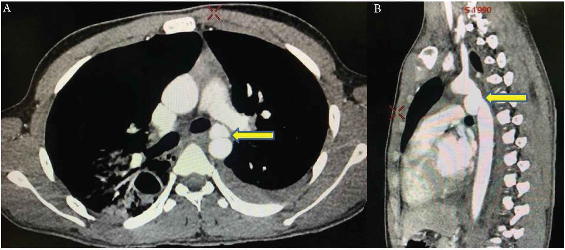
Grade 3 traumatic aortic dissection. (A) Axial Computerized Tomography Angiography (CTA) with intimal flap causing false and true lumen. (B) Coronal CTA revealed pseudoaneurysm (yellow arrows).
On examination, the patient was conscious (Glasgow coma scale 15), with blood pressure 150/90 mmHg, heart rate 75 bpm, respiratory rate 25 breaths/min, oxygen saturation 97% in room air, and axillary temperature 37.1°C.
The patient was unable to move his legs or empty his bladder, and he had numbness distal to T8–T10. All essential blood tests were done on the patient, and their results were within normal limits, apart from increased serum creatinine of 130 µmol/L and blood urea of 10 mmol/L.
The neurologic examination demonstrated that the patient had bilateral flaccid paraplegia (0/5 motor power and hypotonia in both lower limbs) but with preserved motor function in his arms and upper body and no cranial nerve involvement. He had loss of temperature and pain sensation, with a vague numbness from his upper abdomen to the tips of his toes, preserved position and vibratory sensation, and areflexia in both lower limbs. The patient was reviewed by different specialties and nothing else relevant in systemic clinical examination.
The neurology consultation suggested the possibility of ischemic spinal injury with ischemic myelopathy, based on the clinical picture of paralysis secondary to loss of corticospinal tract function with a sensory change from impaired spinothalamic function, as well as signs of infarction of the anterior horn cells, and the very preserved posterior column function. Although the onset of anterior spinal artery syndrome is usually sudden, it may occasionally be gradual (over hours or days) and therefore, an urgent vascular surgery consultation was made.
Results of examination showed pale and cold lower limbs with very weak femoral arteries pulses. CTA was performed rapidly and showed collapsed distal 2 cm of the previous aortic stent graft, because of distal stent struts fracture, with an intimal injury-causing subintimal hematoma and thrombosis and subsequently impaired flow (Figure 2).
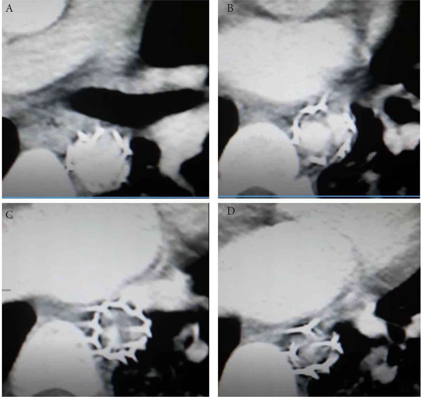
The patient’s Computerized Tomography Angiography (CTA) showing (A, B) previous aortic stent graft distal fracture with intimal injury causing subintimal hematoma and thrombosis, (C, D) collapsed distal 2 cm of the stent.
Next, the patient underwent conventional angiography, under local anesthesia and sedation, through 6F right radial access sheath, which confirmed the diagnosis and the true lumen of the aorta (Figure 3).
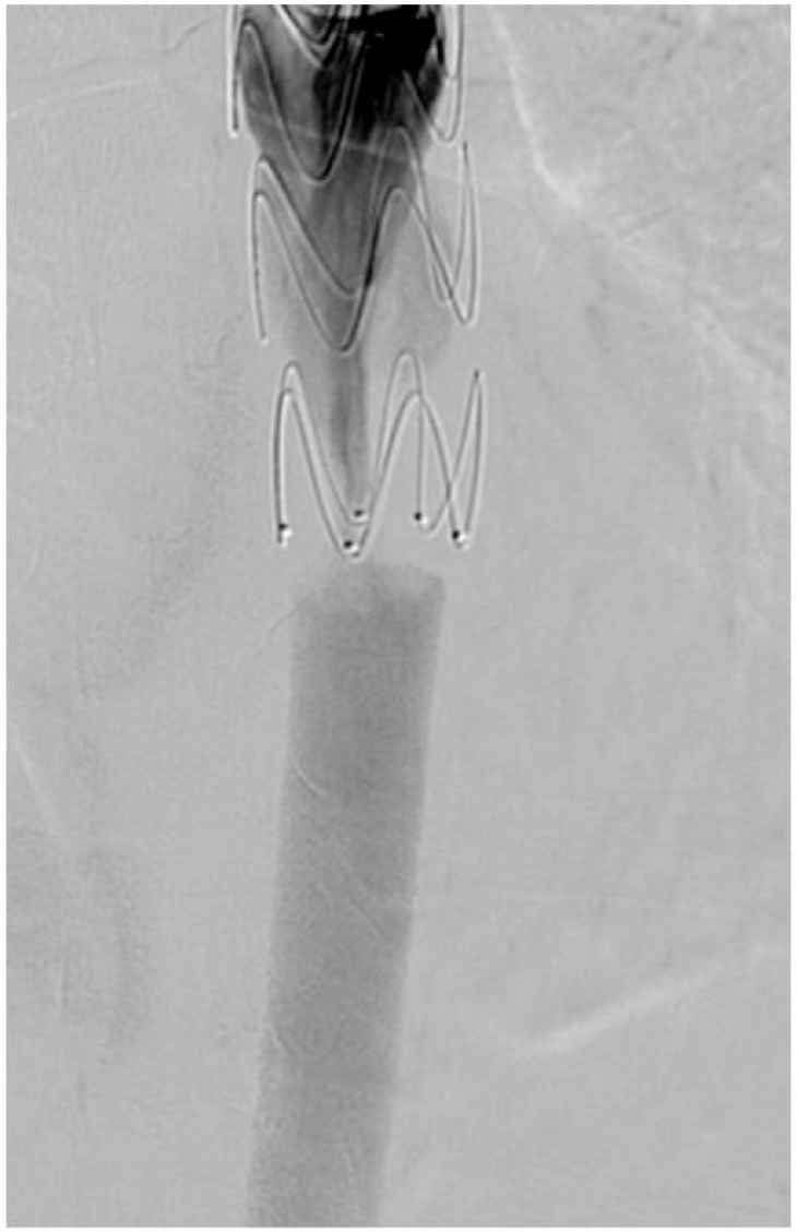
Conventional angiography showing impaired flow at the collapsed distal previous aortic stent graft.
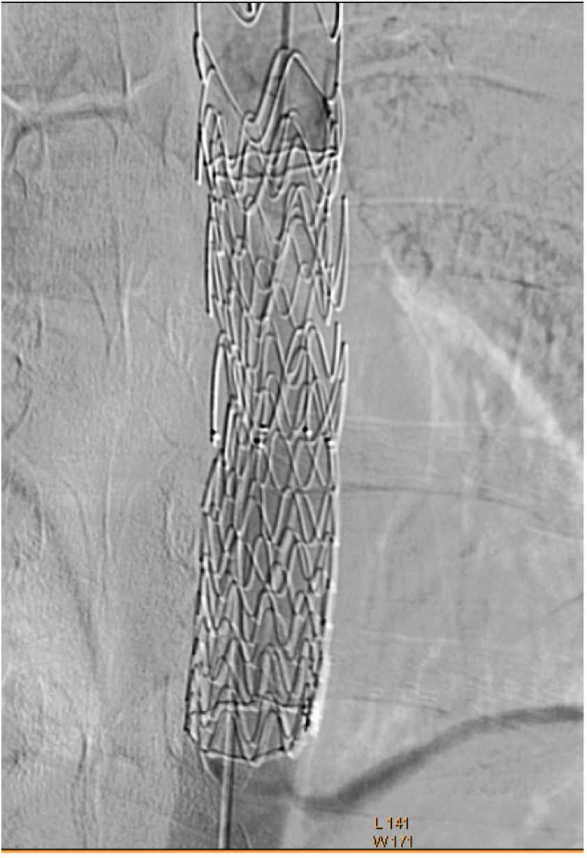
Intervention with right femoral (20 F) accesses sheath, the Gore Covered (Gore & Associates, Flagstaff, AZ, USA) Gore coered stent was successfully deployed with 4 cm overlap with the old stent. Post-deployment dilatation, using a trilobal balloon, was used (Figure 4), and the normal blood flow in the aorta was achieved (Figure 5). Then, the femoral access site was closed using a closure device.
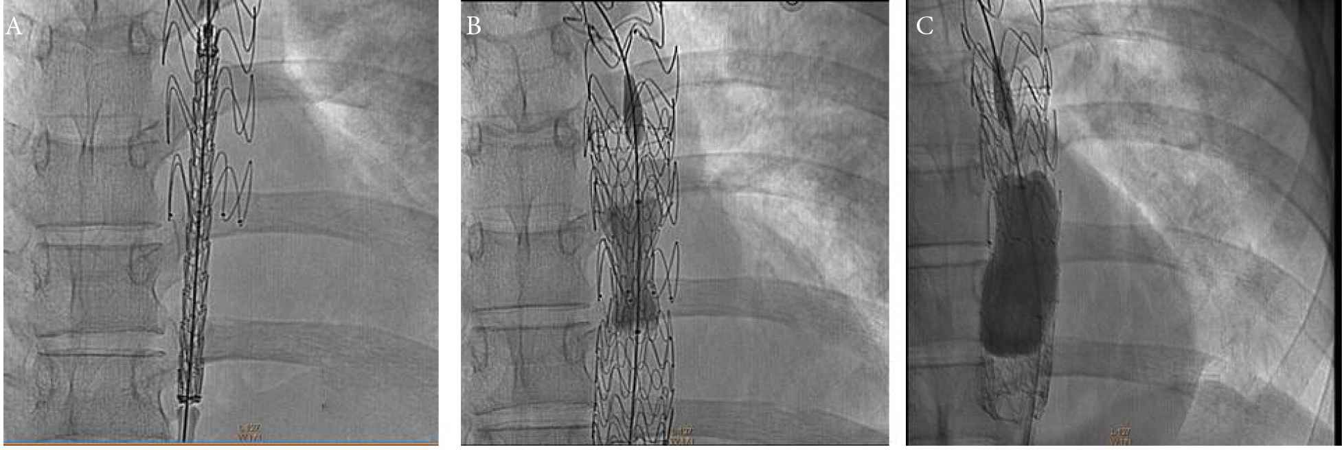
Conventional angiography showing (A) new stent deployment with overlap of the old fractured stent, (B, C) post-deployment dilatation using trilobal baloon.
Restoring the flow after the procedure.
The patient was shifted to intensive care for 1 day, and then, being hemodynamically stable, transferred to the ward after 3 days. The neurology and physiotherapy departments took over for the management of the patient’s deficits. After the intervention, the lower chest and abdominal pain disappeared completely, and intestinal ischemia was excluded but there was no immediate change for the lower limb neurological function.
There was short-term follow-up after the patient was discharged home, lasting nearly 3 months and mainly with the neurology clinic. Unfortunately, as the patient returned to his home country, we were unable to follow him up in his own country for two times.
3. DISCUSSION
Blunt thoracic aortic injury may be fatal if not diagnosed and treated early; in fact, it remains a leading cause of trauma deaths [2,3]. In 1994, Dake et al. [23] reported preliminary results indicating that endovascular stent graft repair is safe in highly selected patients with descending thoracic aortic aneurysms. Meanwhile, in 1997, Semba et al. [24] indicated that stent graft repair is technically possible in acute descending thoracic aortic rupture. Since then, TEVAR has become one of the primary lines of treatment for aortic pathology, and its role now is more prominent in the management of BTAI because of its lower mortality and morbidity as compared to repair by open surgery. Moreover, this procedure has been adopted as a standard of care as a minimally invasive intervention and because of the superior short-term outcomes relative to open surgery [25].
Although early complications are more common in open repair, late complications are more common in endovascular repair [3]. Infection, access site complications, endoleak, stent migration, pseudo-coarctation, and graft occlusion can occur acutely and may require re-intervention or, in rare cases, open repair. Stroke (embolization from wire, device, and/or catheter manipulation in the aortic arch), vertebrobasilar insufficiency (caused by partial or complete coverage of the left subclavian artery), left upper extremity ischemia, and paraplegia/paraparesis are all known but rare acute complications of TEVAR for BTAI with spinal cord injury being the most feared [3].
There are still concerns about the long-term device-related complications of TEVAR in terms of the durability of the stent and inadequate stent graft characteristics, as well as concerns about the progressive aortic expansion with aging and about the age limit to be used in children and adolescents, although it has been reportedly used in patients aged 15 and 8 years [26,27]. Furthermore, the effect of radiation exposure in long-term follow-up from repeated CT scans is another concern with the potential for being Victims of Modern Imaging Technology, mainly in younger patients [25,28].
The importance of our case report comes from the late fracture collapse of the stent, as paraplegia due to spinal cord ischemia occurs in up to 12% of cases after TEVAR [29]. However, the symptoms of spinal cord ischemia typically develop within 12 h after repair [19], and endograft collapse typically occurs within the first 30 days after the procedure, with a median time to collapse of 15 days, and the patient usually presents with clinical symptoms of acute aortic occlusion [30]. It should be noted, that our patient presented more than a year after he had undergone TEVAR.
Long-term success for thoracic aorta endograft placement warrants a device design that maintains stent integrity and durability, and can withstand changes induced by aortic remodeling as the thoracic aorta provides a hostile environment for thoracic stent-graft placement because of its high flow pulsation [21]. Also, oversizing and twisting injury are claimed to be the reasons for stent collapse mainly in adolescent and younger patients, as the growth of the aorta is about 1.5 mm per decade, which can lead to a considerable increase of the aortic diameter from adolescent to later age [31]. Although metal fracture is known to happen as a result of metal fatigue created by the circumferential, radial, and torsional stresses as endografts are subjected to repetitive aortic pulsation [21], this usually occurs earlier. Thus, we believe that strenuous work, including heavy lifting and jumping off scaffolds (as our patient used to do at work), might provoke this result leading to symptoms described above and stent collapse, as it has been reported that twisting and buckling can lead to stent fracture and other related complications [32]. Moreover, most late complications can be repaired with secondary endovascular interventions if recognized with diligent and prompt postoperative surveillance [21].
4. CONCLUSION
Thoracic endovascular aortic repair in adolescent and younger patients warrant further studies and long-term follow-up because of the potential early and late complications.
Strenuous activities of patients after TEVAR and the effect of oversizing of the stent in young adults may contribute to the development of late complications.
CONFLICTS OF INTEREST
The authors declare they have no conflicts of interest.
AUTHORS’ CONTRIBUTION
HAL and ASMN contributed to study conceptualization, writing (review & editing) and formal analysis of the manuscript.
FUNDING
There was no funding for this work and none of the authors had received any financial income/payment from commercial sources, the interests of which are relevant to this research activity.
Footnotes
REFERENCES
Cite this article
TY  - JOUR
AU  - Hussein Ahmed Lateef
AU  - Ahmed Sameer Mahmud Nadeem
AU  - Mutaz I.K. Fakhry Al-Khateeb
AU  - Adel Aswad
PY  - 2021
DA  - 2021/08/16
TI  - Paraplegia Due to Stent Fracture, after Thoracic Endovascular Aortic Repair for Blunt Thoracic Aortic Injury: A Case Report and Review of Literature
JO  - Dr. Sulaiman Al Habib Medical Journal
SP  - 93
EP  - 98
VL  - 3
IS  - 3
SN  - 2590-3349
UR  - https://doi.org/10.2991/dsahmj.k.210811.001
DO  - 10.2991/dsahmj.k.210811.001
ID  - Lateef2021
ER  -