
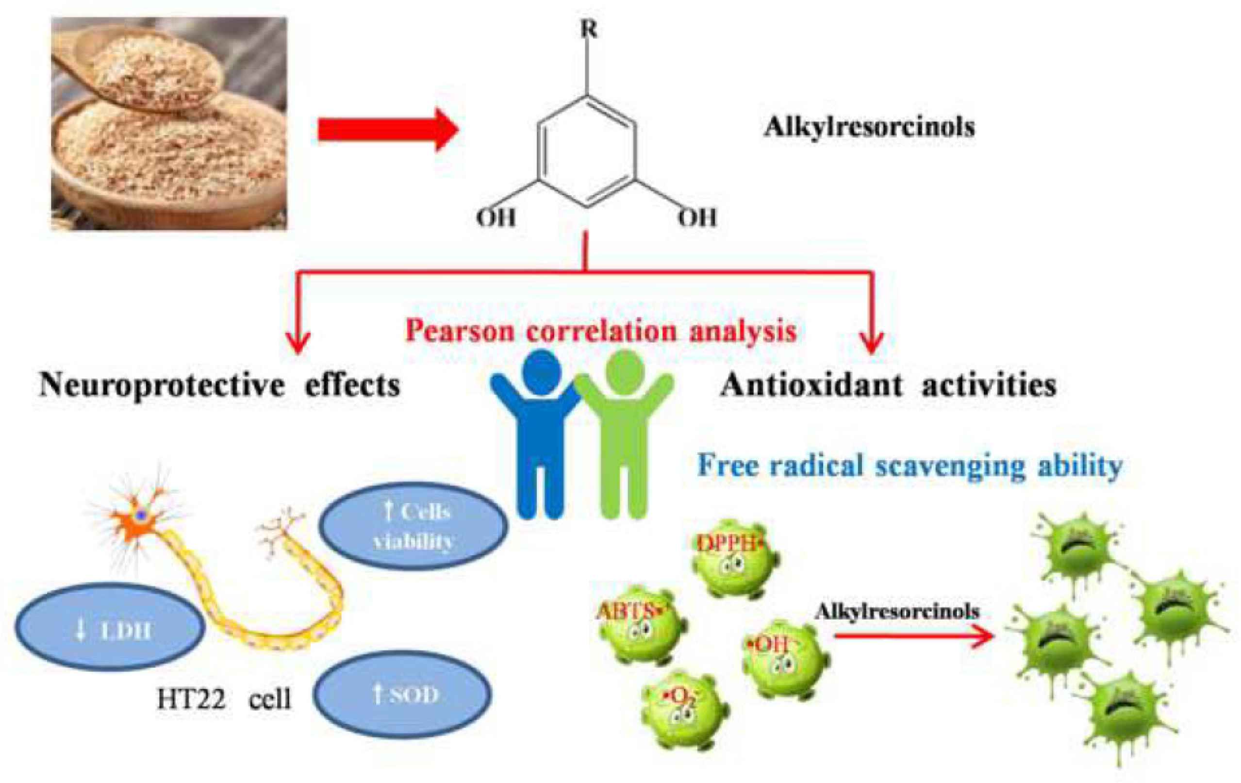
Neuroprotective Effect of Alkylresorcinols from Wheat Bran in HT22 Cells: Correlation with in vitro Antioxidant Activity
- DOI
- 10.2991/efood.k.210125.001How to use a DOI?
- Keywords
- Wheat bran; alkylresorcinols; neuroprotective effect; antioxidation
- Abstract
Alkylresorcinols (ARs), which are phenolic lipids found in wheat bran, have attracted a considerable amount of attention because of their antioxidant properties. However, the mechanism of ARs for regulating neuroprotective activity remains unclear. The correlation between the cellular neuroprotective effects of ARs in HT22 cells and their in vitro antioxidant activity were investigated. The results showed that two main constituents of crude ARs were C19:0 and C21:0, which was identified by high-performance liquid chromatography-atmospheric pressure chemical ionization tandem mass spectrometry. Pre-treatment with 400 μg/mL crude ARs could protect from H2O2-induced cellular damage involving low cell viability, leakage of lactate dehydrogenase and decreased superoxide dismutase activity. The maximum radical scavenging ability of DPPH•, ABTS•+, •
- Graphical Abstract
- Copyright
- © 2021 The Authors. Publishing services by Atlantis Press International B.V.
- Open Access
- This is an open access article distributed under the CC BY-NC 4.0 license (http://creativecommons.org/licenses/by-nc/4.0/).
1. INTRODUCTION
Alkylresorcinols (ARs) are one of the major phenolic compounds in whole grain cereals; they account for 0.015–0.3% of the grains dry weight [1]. ARs are special phenolic lipids in the outer layers of wheat and rye grains and other cereal grains [2] and have been considered biomarkers of whole cereals grains intake [3–5]. The homologs of ARs were distinguished by their alkyl side chain, which vary from 17 to 25 carbon atoms [6]. In previous research, High-performance Liquid Chromatography Mass Spectrometry (HPLC-MS) was extensively performed to measure and identify individual ARs homologs in cereals products. Evidence showed that the majority of homologs was saturated; only 0.5–20% unsaturated and oxygenated homologs were detected in cereals [7]. The chain length and structure of AR homologs have significant impacts on their bioactivity [8].
Polyphenols and phenolic compounds are bioactivity nutrients that generally originate from fruits, vegetables and cereals [9]. In recent years, numerous experimental researches demonstrated that polyphenols and phenolic compounds were beneficial for neurodegenerative diseases [10,11]. The neuroprotective activity of phenols may be closely related to their antioxidant activity. For example, epigallocatechin-3-gallate, the main polyphenol constituent in green tea, could be used as neuroprotective agents because of its radical scavenging capacity, iron-chelating property and antioxidant enzymes regulation ability [12]. Shi et al. [13] discovered that isoliquiritigenin could deduce reactive oxygen species (ROS) production to prevent cell apoptosis and exhibit cellular neuroprotective and antioxidant activity in H2O2-treated PC12 cells. ARs are potential natural antioxidants, which have a similar straight aliphatic hydrocarbon side chain and single phenolic ring with tocopherols. Moreover, ARs could prevent lipid peroxidation in natural membranes, which effectively protected the erythrocyte membrane from H2O2-induced oxidation [14]. Numerous studies indicated that ARs have many bioactivities, which could reduce blood cholesterol and prevent muscle atrophy and oxidative damage [15–17]. However, relevant studies on the neuroprotective effect of ARs are limited.
Oxidative stress has been regarded as the main pathological factor in the initiation and development of neurodegenerative diseases [18]. Oxidative stress may induce membrane damage in neural cells and accelerate the development of neurodegenerative disorders [19]. Evidence indicated some relationships between neuroprotective effects and antioxidant activity. Phenolic compounds were able to protect neurons against oxidative damage owing to their antioxidant properties [20]. Lu et al. [21] discovered that gallic acid could cross through the liposome membrane to react with the free radical and exhibited neuroprotective effect. ARs have been regarded as antioxidant phenolic lipids, which were effective antioxidants in phospholipid bilayers and could prevent membrane lipid peroxidation. Therefore, ARs may have neuroprotective effects because of their antioxidant activity. However, the correlation between neuroprotective effects and antioxidant activity of ARs has never been determined.
Therefore, the objectives of this study were (1) to obtain ARs with ultrasound from wheat bran and identify ARs homologs by HPLC-MS, (2) to explore the neuroprotective effects and antioxidant activity of ARs in vitro, and (3) to evaluate the relationship between the neuroprotective effects of ARs and the antioxidant activity of ARs in vitro by Pearson correlation analysis.
2. MATERIALS AND METHODS
2.1. Materials and Reagent
Wheat bran was collected from the Lingshan grain depot (Jiangsu, China). The moisture of all wheat bran was less than 14%. Wheat bran was milled to flour, passed through an 80-mesh sieve, and then stored at −20°C until analysis. Dulbecco’s Minimal Essential Medium (DMEM), Fetal Bovine Serum (FBS), penicillin and streptomycin, Methylthiazolyl Tetrazolium (MTT), Dimethyl Sulfoxide (DMSO) and H2O2 solution (30%, w/w) were purchased from Sloarbio Technologies (Beijing, China). Lactate Dehydrogenase (LDH), Superoxide Dismutase (SOD) and Bicinchoninic Acid (BCA) were purchased from the Beyotime Biotechnology Institute (Nanjing, China).
2.2. Preparation of Crude ARs from Wheat Bran
Wheat bran ARs was obtained by method described in previous studies with some modifications [22]. Crude ARs were extracted by ethyl acetate coupled with an ultrasonic treatment. The mixture was centrifuged and the supernatant fraction was collected and filtered. Ethyl acetate was removed in vacuo at 45°C to obtain crude ARs extracts. The main composition of crude ARs extracts were determined by HPLC-Atmospheric Pressure Chemical Ionization Tandem Mass Spectrometry (HPLC-APCI-MS/MS).
2.3. Main Components Analysis of Crude ARs by HPLC-APCI-MS/MS
Alkylresorcinols was identified according to a previously reported method with some modification [23]. The quantitative analysis of ARs was analyzed by ZORBAX Stable Bond-C18 column (150 × 4.6 mm2, 5 μm, Agilent Technologies, California, USA) via HPLC at 35°C. The column was eluted with mobile phase A [0.1% (v/v) formic acid in methanol] and mobile phase B [0.1% (v/v) formic acid in water] at a flow rate of 0.5 mL/min with 20 μL injections. ARs were monitored with a UV detector at 280 nm. The elution procedure started with 85% of eluent B and increased to 90% of eluent B from 0 to 5 min, gradually increased to 95% of eluent B in 10 min, then increased to 100% in 15 min, and remained at 100% of eluent B from 15 to 35 min. The recording and evaluation of the chromatograms were performed with Agilent Chem station software (version C.01.03, Agilent Technologies).
The MS qualitative analyses of ARs were performed by mass spectrometer equipped with an APCI source and Agilent Chem Station software. APCI/MS conditions were optimized as follows: positive ion mode; drying gas (N2), 9 mL/min; nebulizer pressure 60 pis; drying gas temperature, 350°C; capillary voltage 3500 V; and positive-ion mass range (m/z) 100–800.
2.4. HT22 Cellular Activity Experiment
2.4.1. Cell culture
The mouse hippocampal neuronal cell line (HT22 cells) was obtained from the Institute of Biochemistry and Cell Biology (CAS, Shanghai, China). HT22 cells were cultured in DMEM, including 10% FBS and 1% streptomycin/penicillin, and incubated in a 37°C cell culture incubator with 5% CO2. HT22 cells were seeded in well plates. After 24 h, the medium was discarded and incubated with 100 μL fresh DMEM that contains various concentrations (0, 50, 100, 200, 400 and 800 μg/mL) of ARs for 24 h. Trolox (50 μmol/L) was treated as the positive group. Then, the cells were treated with H2O2 (200 μmol/L) for 2 h to induce oxidative damage.
2.4.2. Cell cytotoxicity activity, cell viability and morphology
HT22 cells were seeded in 96-well plates and each well contains 1 × 10⁴ cells. The MTT assay was utilized to evaluate the cytotoxicity and cell viability of HT22 cells. MTT solution (20 μL, 5 mg/mL) were added to each well for 4 h. Subsequently, 100 μL DMSO was added to plates for 30 min at 37°C. After blue formazan crystal completely solubilized, the absorbance at 570 nm was measured by using a microplate reader (Spectra MAX 190, Molecular Devices Inc., CA, USA). The cell viability of the untreated cells group was considered as 100%. To observe the morphological changes, the morphology of HT22 cells were captured using inverted phase contrast microscopy.
2.4.3. LDH and SOD assay
HT22 cells (1 × 10⁴ cells) were seeded in 96-well plates and cell supernatants was applied to evaluate the leakage of cytoplasmic LDH enzyme. LDH leakage was measured according to the manufacturer’s instructions. 1 × 10⁵ cells/mL were seeded in a 6-well plate and the intracellular SOD levels of HT22 cells were detected by an SOD and BCA kit. The intracellular SOD activity and protein concentrations were determined according to the manufacturer’s instructions. The absorbance of LDH and SOD was determined by a microplate reader at 490 and 450 nm.
2.5. Assay of Antioxidant Activity of ARs in vitro
2.5.1. DPPH• radical scavenging ability
The DPPH• free radical scavenging activity of ARs were determined according to the reported method with slight modifications [24]. ARs were homogeneously dispersed with analytical grade methanol to prepare different concentrations samples (0.125–2.0 mg/mL). In addition, 50 μL samples and 200 μL DPPH solution (0.4 mmol/L) were added and mixed. After the mixture incubated for 30 min in the dark at room temperature, the absorbance was measured at 517 nm by a microplate reader. Trolox was used as the positive control. The DPPH scavenging ability was calculated by the following equation:
2.5.2. ABTS•+ radical scavenging ability
The ABTS•+ radical scavenging activity assay described by Liu et al. [25] was utilized with some modifications. The 7 mmol/L ABTS stock water solution was mixed with 2.45 mmol/L K2S2O8 and placed in the dark at room temperature for 12 h to generate ABTS•+. The solution was diluted with PBS buffer solution (0.2 mol/L, pH 7.4) and measured at 734 nm by a microplate reader until the absorbance decreased to approximately 0.7 to obtain a working solution. After 20 μL sample (0.125–2.0 mg/mL) was mixed with 200 μL working solution, the absorbance was measured at 734 nm. Trolox was used as the positive control. The ABTS•+ scavenging ability was calculated by the following equation:
2.5.3. •
O 2 −
•
2.5.4. •OH– radicals scavenging activity
The •OH– radical scavenging activity was determined according to the reported method [27]. The reaction mixture contained 0.2 mL ferrosin (0.75 mmol/L), 0.3 mL phosphate buffer (0.15 mol/L, pH 7.4), 0.2 mL FeSO4 (0.75 mmol/L) and 0.2 mL sample solutions (0.125–2.0 mg/mL). To terminate the reactions, 0.2 mL H2O2 (0.01%, w/v) was added. After incubation at 37°C for 60 min, the absorbance was measured at 536 nm. Trolox was utilized as the positive control. The •OH– scavenging activity was calculated by the following equation:
2.6. Statistical Analysis
Data was expressed as the mean ± Standard Deviation (SD) in triplicates. Origin 8.5 (Origin Lab, USA) was selected for the statistical and graphical analysis. A correlation analysis was performed using the Pearson correlation coefficient. SPSS 22.0 (IBM Corp., Armonk, NY, USA) was conducted to determine the variations between and within experimental groups; the statistical significance was set to p < 0.05 or p < 0.01.
3. RESULTS AND DISCUSSION
3.1. Main Structural Composition of Crude ARs
In our study, the highest extractive rate could be 89.27% when crude ARs were isolated and extracted by ultrasound-assisted ethyl acetate extraction. As shown in Figure 1A, many peaks were observed in crude ARs. Peak 1 and 2 were selected for the subsequent analysis. Peak 1 and 2 were characterized as C19:0 and C21:0 by MS spectra analysis (Figure 1B and 1C), which is consistent with previous literature [28,29]. The same MS2 product ion was found in both Peak 1 and 2 at m/z 123.4. Apart from m/z 123.4, m/z 111.4 and m/z 141.5 was another MS2 product ion in Peak 1 and 2, respectively.
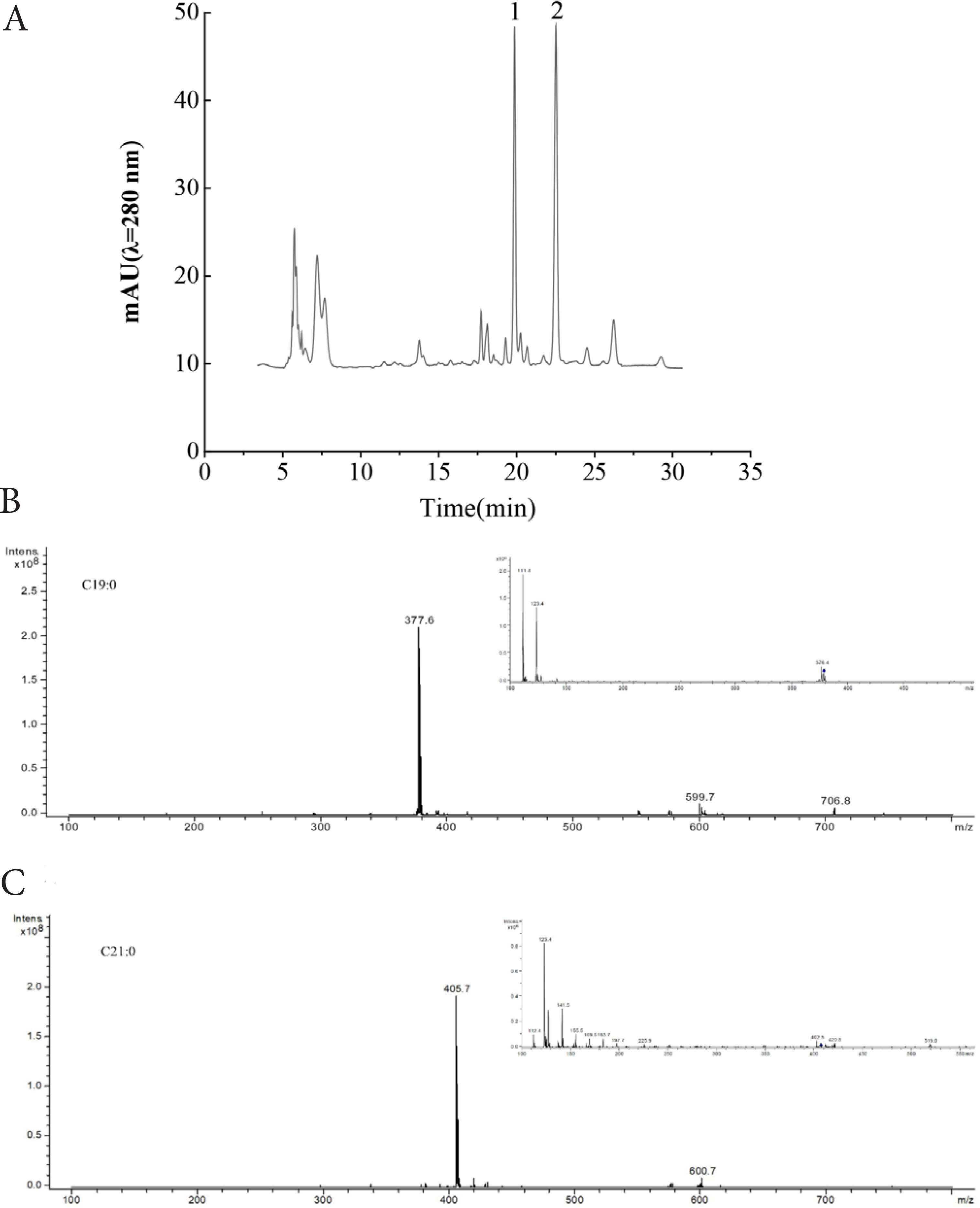
Chromatogram of wheat bran crude ARs. (A), HPLC chromatogram; (B) and (C), ion chromatograms analysis of two main ARs homologs compounds by HPLC-APCI-MS. Peak 1 and 2 (A) are two main ARs homologs and correspond to (B) and (C), respectively.
3.2. Evaluation of Cytotoxicity Activity and Cell Viability of ARs in H2O2-Induced HT22 Cells
As shown in Figure 2A, ARs had different effects on cellular viability when the concentrations ranged from 50 to 800 μg/mL. When the concentration increased to 800 μg/mL, the cellular viability significantly decreased, which indicates that ARs generated cytotoxicity to HT22 cells. Therefore, the concentrations ranged from 50 to 400 μg/mL were chosen for subsequent experiments.
In this experiment, Trolox (50 μmol/L) was used as a positive control due to its antioxidative effects [30]. After treatment with H2O2 (200 μmol/L) for 2 h, the cell viability of HT22 cells significantly decreased to 47.28% (Figure 2B). However, the cell viability increased from 53.56% to 84.35% in the cells experiment groups with a dose-dependent manner. Among all the concentrations, 400 μg/mL ARs showed the most significant effect to improve cell viability. The results indicated that ARs have significant protective effects for alleviating the H2O2-induced oxidative injury in HT22 cells.
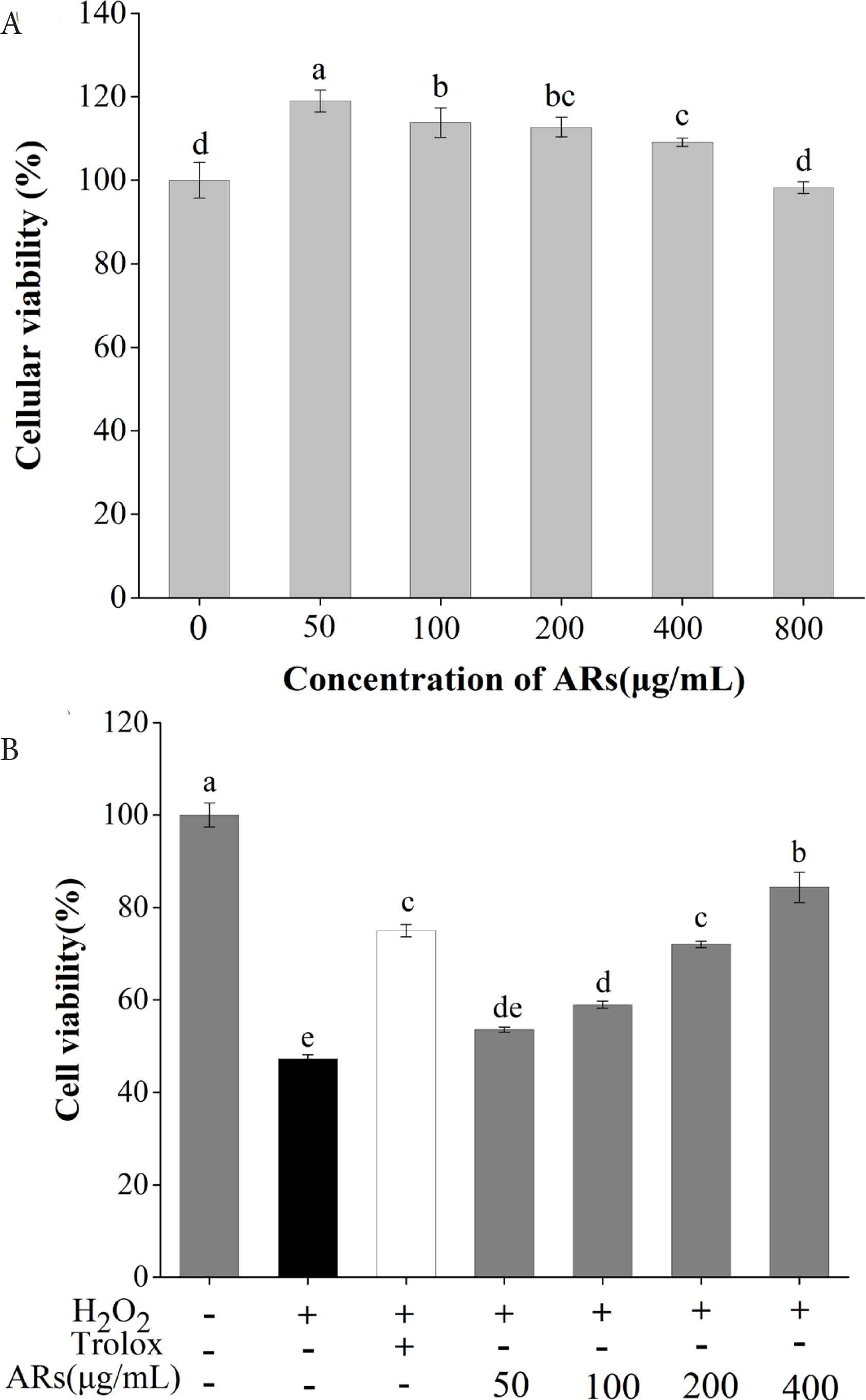
Effects of crude ARs on HT22 cell viability. (A) Determination of ARs dose; (B) Protective effect of ARs in H2O2-damaged HT22 cells. Results marked with different letters represent significant differences (p < 0.05).
3.3. Effect of ARs on LDH Release and Intracellular SOD Activity in H2O2-Induced HT22 Cells
As shown in Figure 3A, the percentage of LDH release degree remarkably increased to 176.15% when cells were treated with H2O2. H2O2 can induce oxidative stress, which cause LDH leakage into the culture medium and break down the integrity of cell membranes [31]. All these values clearly implied that ARs could decrease the LDH release degree. 400 μg/mL ARs reduced the percentage of LDH leakage to 119.78%. Therefore, ARs effectively attenuated the levels of extracellular LDH and avoid the cell membrane fracture induced by H2O2.
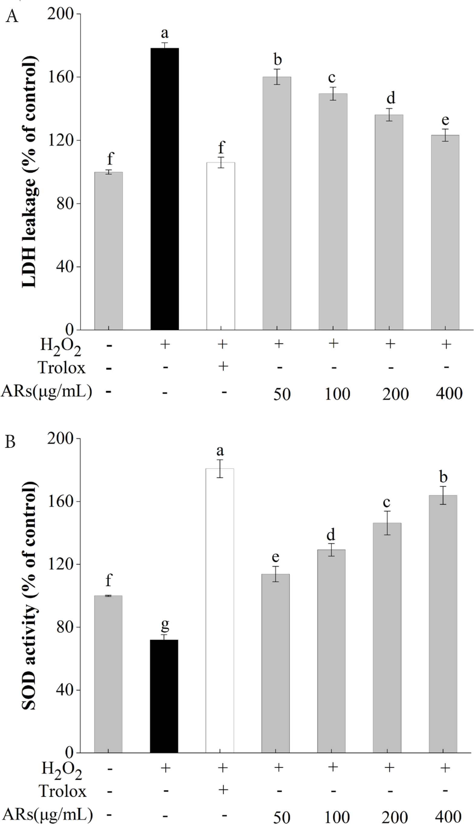
Effect of ARs on intracellular LDH leakage rate (A) and SOD activity (B) in H2O2-induced HT22 cells. Results marked with different letters represent significant differences (p < 0.05).
The effect of ARs on the intracellular SOD activity in H2O2-induced HT22 cells is shown in Figure 3B. The results showed that the intracellular SOD activity was markedly lower after exposure to H2O2. However, ARs could increase the intracellular SOD activity in a dose-dependent manner compared with the control group. When the concentration of ARs increased to 200 μg/mL, a significant effect on enhancing the intracellular SOD activity was observed. SOD is a pivotal intracellular antioxidant enzyme that could prevent oxidative stress induced by H2O2 and has an essential role in defense mechanisms against cell-death [24]. Thus, the results showed that ARs could alleviate H2O2-caused oxidative damage by improving the activity of SOD enzyme.
3.4. Antioxidant Activity of ARs in vitro
The antioxidant activities of ARs are shown in Figure 4. With an increase in the AR concentration, the in vitro antioxidant activity enhanced. The DPPH radical scavenging ability of ARs were 10.62–85.45%, respectively. The maximum ABTS•+, •
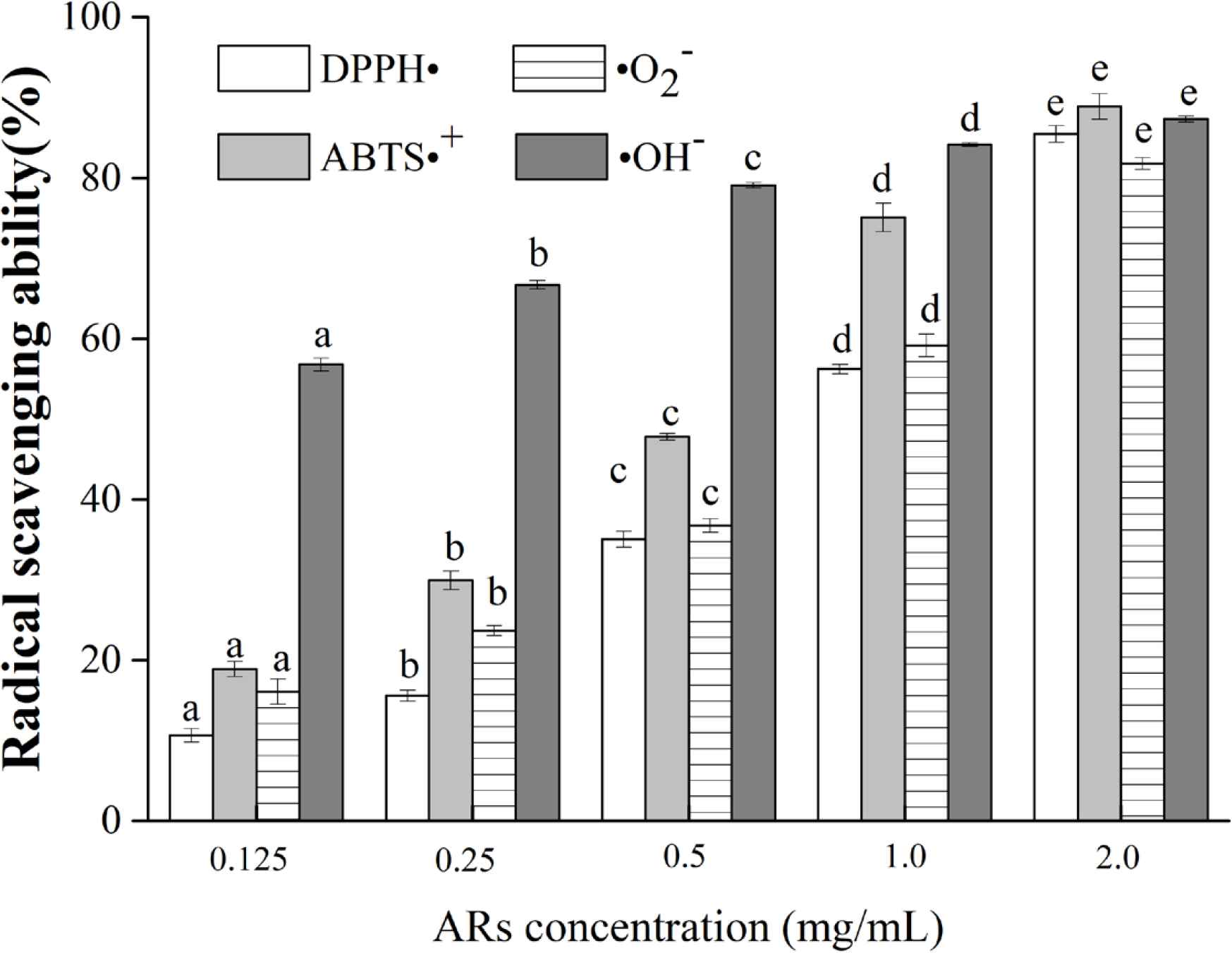
DPPH•, ABTS•+, •
3.5. Correlation Analysis between Cellular Neuroprotection and in vitro Antioxidant Activity
As shown in Figure 4, with an increase in the AR concentration, its antioxidant activity enhanced in vitro. Table 1 showed the coefficient among various parameters, the LDH release rate had a negative correlation with in vitro antioxidant activity. The value of the coefficient between •
| Cell viability | LDH | SOD | |
|---|---|---|---|
| DPPH• | 0.9955** | –0.9971** | 0.9972** |
| ABTS•+ | 0.9776* | –0.9927** | 0.9918** |
| •
|
0.9988** | –0.9974** | 0.9960** |
| •OH– | 0.8909 | –0.9384 | 0.9439 |
Indicates significant differences at p < 0.05 and
indicates very significant differences at p < 0.01 by Pearson correlation analysis.
Correlation coefficient between cellular neuroprotective effect and in vitro antioxidant activity of ARs in HT22 cell
The ABTS•+ radical scavenging activity showed significant correlation to cell viability and correlated extremely significantly with LDH release rate and intracellular SOD level, marked with correlation coefficients 0.9776, –0.9927 and 0.9918. The DPPH• and •
Alkylresorcinols homologs are amphiphilic phenolic lipids that are detected in wheat, rye and other wheat [34]. Hydrophobicity is a particularly important factor in evaluating the intracellular antioxidant capacity [21]. Hydrophobic antioxidants are generally more effective for preventing oxidative stress-induced neurodegenerative diseases. On the other hand, lipophilic polyphenols have a better membrane-penetrating capacity, which may facilitate they enter into neuron cells. Moreover, the human brain has a large content of lipids, where lipophilic antioxidants may represent a higher neuroprotective effect on the oxidative stress [35]. Therefore, the special amphiphilic characteristic account for neuroprotective effects of ARs.
Additionally, the aliphatic side chains of ARs participated in their antioxidant properties [36]. After HT22 cells were exposed to H2O2, the membrane became severely damaged and generated a large content of reactive oxygen species. The alkyl tail of ARs would interact with phospholipids or proteins to regulate the phospholipid bilayer properties and physicochemicals of the membrane and then modulate the activities of some antioxidant enzymes [37]. After ARs crossed the cell membrane and entered the HT22 cells, they would react with the cellular reactive oxygen species and other free radicals. With the formation of hydroxyl radicals, the electron density of the antioxidant system would change, which cause oxidation of the ARs ring to protect HT22 cells from oxidative damages. Thus, the cellular neuroprotective activity of ARs may depend on its antioxidant activity. Korycińska et al. showed that the difference in the alkyl chain length within AR homologs has a slight impact on their antioxidant properties. In this study, we acknowledged that C19:0 and C21:0 were the predominant ARs homologs in wheat bran. Therefore, the findings concluded that the cellular neuroprotective activity had a positive correlation with the in vitro antioxidant activity. ARs can be used as neuroprotective agents and future research is warranted.
4. CONCLUSION
Wheat bran ARs possess not only antioxidant properties but also neuroprotective activity. The neuroprotective activity of ARs in HT22 cells was correlated with its antioxidant activity in vitro. C19:0 and C21:0 are two major ARs homologs in wheat bran and could be the key factors that relate to the •
CONFLICTS OF INTEREST
The authors declare they have no conflicts of interest.
AUTHORS’ CONTRIBUTION
YZ carried out the experiments and interpreted the results. FF did some data analysis work. YF designed the study and interpreted the results. PL, JX, XS, QL and QH instructed the instrument operation.
ACKNOWLEDGMENTS
This work was supported by the National Key Research and Development Program of China (2016YFD0401203), Priority Academic Program Development of Jiangsu Higher Education Institutions (PAPD), and National Youth Talent in Grain Area Support Program of China (LQ2018301).
Footnotes
REFERENCES
Cite this article
TY  - JOUR
AU  - Yanyu Zou
AU  - Fengjiao Fan
AU  - Yong Fang
AU  - Peng Li
AU  - Ji Xia
AU  - Xinchun Shen
AU  - Qin Liu
AU  - Qiuhui Hu
PY  - 2021
DA  - 2021/01/31
TI  - Neuroprotective Effect of Alkylresorcinols from Wheat Bran in HT22 Cells: Correlation with in vitro Antioxidant Activity
JO  - eFood
SP  - 13
EP  - 20
VL  - 2
IS  - 1
SN  - 2666-3066
UR  - https://doi.org/10.2991/efood.k.210125.001
DO  - 10.2991/efood.k.210125.001
ID  - Zou2021
ER  -