
Perspectives for Recognition and Rapid Detection of Foodborne Pathogenic Bacteria Based on Electrochemical Sensors
- DOI
- 10.2991/efood.k.210621.001How to use a DOI?
- Keywords
- Food safety; foodborne pathogenic bacteria; recognition; rapid detection; electrochemical sensor; signal amplification
- Abstract
Due to the misuse of pathogen-infected food, human beings continue to face serious diseases and even deaths. Thus, researchers from all walks of life concern on prevention, detection, and resistance for pathogenic bacteria. It is necessary to develop and explore a fast, cost-effective, portable, and efficient detection technology for pathogens. Recently, electrochemical sensors have been widely used for food safety analysis, due to their increasingly high sensitivity and selectivity. In particular, researchers have made significant breakthroughs on signal enhancement strategies, quantitative methods, and miniaturization of the instrument, which can be used as a reference to food safety analysis. Moreover, a device integrating facile working procedures, miniaturization, and automation must be indispensable. In order to meet the needs of People’s Daily life, household device and point-of-care (POC) testing need to be combined with electrochemical sensing technology. In this review, the state of the art in electrochemical sensing for recognition and rapid detection of foodborne pathogenic bacteria is summarized from perspectives of common foodborne pathogens, especially focusing on real-time monitoring, lab-on-a-chip, and photoelectrochemical platform applied in food and medical treatment fields. Furthermore, the limitations and prospects of recognition elements, active nanomaterials, and optical materials, that are essential components in sensing systems on future research directions, were evaluated.
- Graphical Abstract
- Copyright
- © 2021 The Authors. Publishing services by Atlantis Press International B.V.
- Open Access
- This is an open access article distributed under the CC BY-NC 4.0 license (http://creativecommons.org/licenses/by-nc/4.0/).
1. INTRODUCTION
Foodborne disease caused by human ingestion of pathogens infected food is the main sources of food safety problems. Pathogenic bacteria that cause foodborne disease are Escherichia coli (E. coli), Salmonella spp., Staphylococcus aureus (S. aureus), Listeria monocytogenes (Lm), Campylobacter spp., Clostridium botulinum, Shiga toxin-producing Escherichia coli and Enterobacter sakazakii [1,2]. The main symptoms of foodborne pathogens on the human body are dysentery, food poisoning, diarrhea and even death [3]. Annually, the number of deaths from bacterial infection is extremely high, with the prediction of 13 million deaths in 2050 all over the world [4]. Especially in the USA, the most commonly reported foodborne pathogenic bacteria are responsible for more than 91% of foodborne outbreaks [5]. Therefore, it is of great significance in recognition and sensitive detection of foodborne pathogenic bacteria.
Conventional culturing methods, immunological analyses, and nucleic acid-based techniques are known as main methods for detection of foodborne pathogenic bacteria. The complicated culturing method is to identify the colonies through biochemical and serological testing under strict culture conditions [6]. Although the culture technique has long been regarded as the gold standard method, its specificity is poorer than that of the antibody-antigen binding based immunoassay [7]. At present, there are some drawbacks in immunological techniques, such as time consumption operation complication (5–6 days) and limited information, namely failing to discriminate species [8–10]. Subsequently, nucleic acid-based techniques relied on deoxyribonucleic acid (DNA) probe have been widely accepted owing to these merits of high specificity and sensitivity. Despite nucleic acid-based methods provide significant application, the presence of inhibitors, misidentification of dead and living cells, expensive equipment and reagents can arise some problems [9]. Overall, traditional methods could not make a timely response to possible risks, even though these technologies have some advantages. Consequently, there is an urgent need for developing and exploring technologies addressing food safety issues.
In recent years, researchers have made some good achievements by fluorescence, chromatography, mass spectrometry, and electrochemical methods to detect pathogens [11–13]. Among these methods, electrochemical methods are among the most promising candidates for the detection of pathogenic bacteria in a low-cost, highly sensitive and selective manner [14]. However, it remains a challenge for rapid and cost-efficient detection of foodborne pathogens. So far, there has been made major breakthroughs in the field of pathogen assays with the emergence of new technologies. Electrochemical collision sensor has been applied to determine live cell viability that can oxidize or reduce redox species to decrease diffusion current of the ultramicroelectrode [15]. The sensor is suitable for monitoring cancerous cells in biological solution and evaluating the validity of antimicrobial agents. In addition, researchers developed a portable sensing platform including antibody capturing cell and impedance analysis unit [16]. The loop-mediated isothermal amplification (LAMP)-based lab-on-a-disc technology could detect a concentration of 1 CFU/mL. In addition to the theoretical and semi-theoretical sensors, the number of POC and commercial kits is undeniably increasing [17,18]. These devices are more inseparable from electrochemical sensing technology [19,20]. Furthermore, innovative, efficient, and commercial equipment must be a durative goal.
Inspired by the content mentioned above, it is intensively expected to develop a miniature, intelligent, integrated, commercial, ultrasensitive, and super-specific device until the realization of full automation. Therefore, this paper reviews the advancements of electrochemical detection for pathogenic bacteria in two parts: (1) Recognition elements and electrochemical methods-based sensors, (2) Electrochemical sensors for detection of various foodborne pathogenic bacteria emphasizing real-time monitoring, microfluidic and automated devices. Moreover, the future research direction and long-term forecast for pathogenic bacteria are discussed.
2. RECOGNITION ELEMENTS AND ELECTROCHEMICAL METHODS-BASED SENSORS
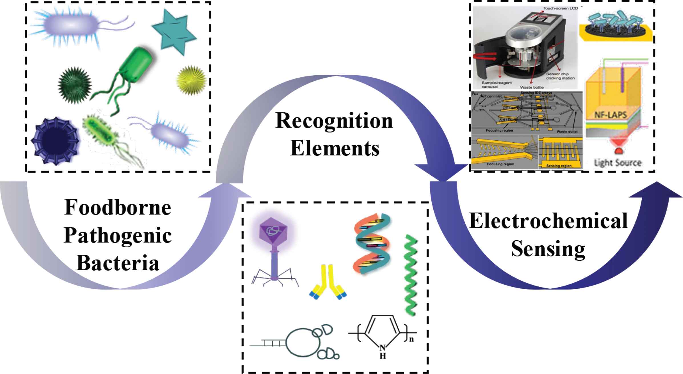
Electrochemical sensor has attracted public attention due to quantitative determination of foodborne pathogen bacteria and prevention of foodborne diseases. One basic electrochemistry sensor includes the recognition elements with high specificity and electrochemical detection methods with high sensitivity [21]. The basic principle of electrochemical sensing pathogen bacteria is presented in Scheme 1. The most important procedure in the preparation of sensors is surface modification including adsorption, self-assembled monolayers (SAM), covalent attachment, and imprinting [4]. The biochemical reaction on the surface of electrode can change the physical parameters such as pH, ion concentrations, oxygen consumption, potential difference, resistance or current. Changes in physical parameters can be represented as electrical signals obtained by electrochemical analysis instruments. These changes are related to the concentration of analytes.
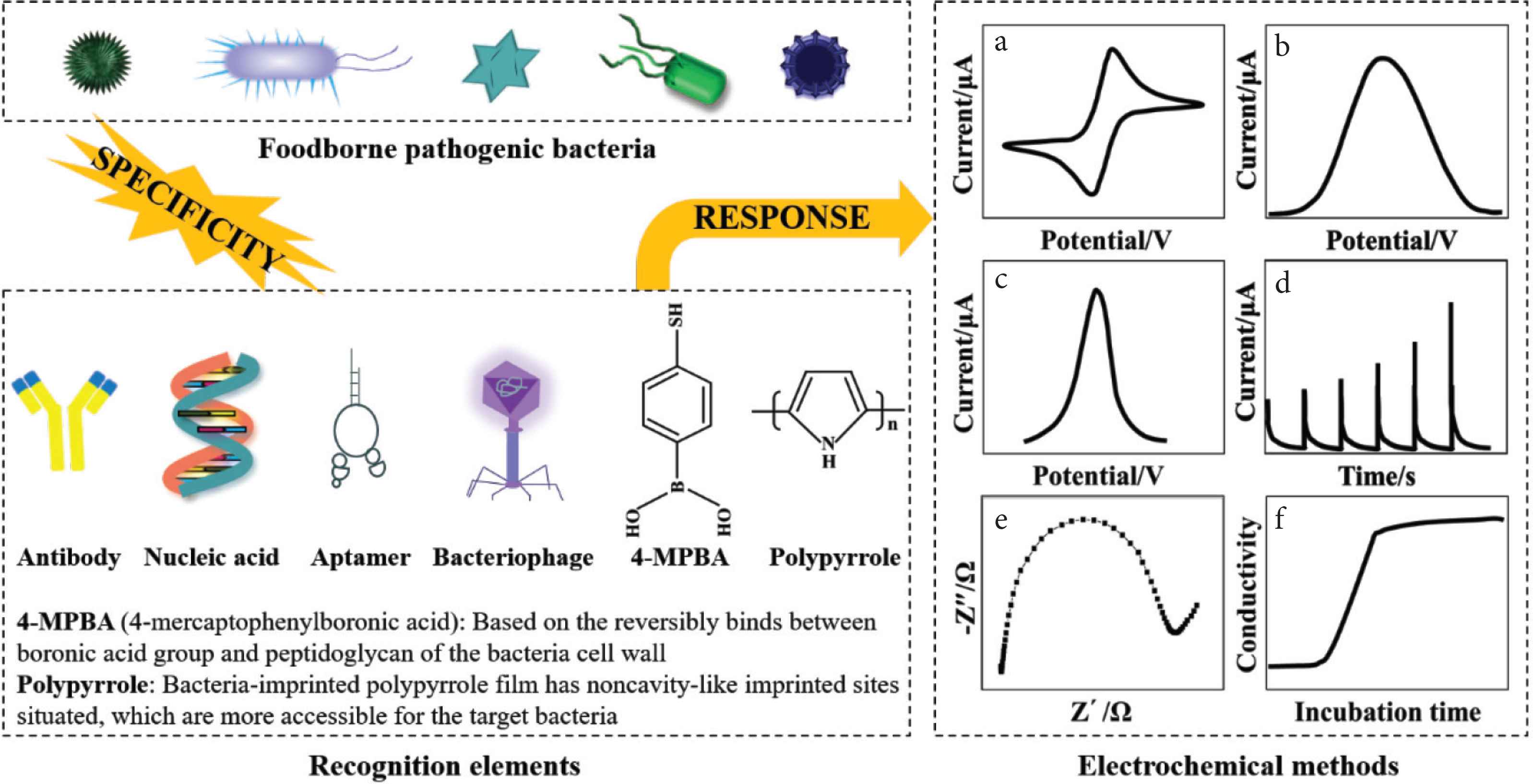
Illustration of electrochemical sensors for recognition and detection of foodborne pathogenic bacteria (a–f: CV, DPV, SWV, amperometry, EIS, conductometry).
As an essential factor to improve specificity of the sensing system, recognition elements mainly consist of purely natural substances and artificial composites. Natural recognition elements include antibodies, DNA probes, enzymes, antimicrobial peptide (AMPs), phages, cells, and tissues. And artificial recognition elements such as aptamers, bacteria-imprinted polymers (BIP) film and synthetic peptides could capture target bacteria [22]. Moreover, BIP film is favored because of its superior electrical conductivity than other bioreceptors. The specific binding of bioreceptor to the pathogen must be independent of the substrate of actual sample.
After the modification of working electrode, electrochemical measurements such as differential pulse voltammetry (DPV), electrical impedance spectroscopy (EIS), amperometry, cyclic voltammetry (CV), square wave voltammetry (SWV), amperometry, and conductometry are used to highly sensitive detection of pathogens. Generally, electrochemical sensing is performed using two (reference and working electrodes) or three electrodes (auxiliary, reference and working electrodes) [22]. To date, various working electrodes such as screen-printed carbon electrodes (SPCE), laser-induced graphene (LIG) electrodes and microarray electrodes have been used to sensitively detect foodborne pathogen bacteria [23–25]. Additionally, the use of triple-helix DNA and amplification reactions also could improve the sensitivity of the sensor [26].
Based on enhancing the sensitivity and selectivity of electrochemical pathogen sensors, researchers have focused on the design of functional integrated platforms and portable devices. Microfluidic device has integrated pretreatment unit, capture unit and detection unit [27,28]. There is no doubt that the use of microfluidic devices simplifies the actual sample pretreatment process. However, current electrochemical pathogen sensors still require pretreatment steps to analyze actual samples, except for juice [29]. So far, a few microfluidic devices have applied for outdoor analysis and clinical diagnosis [30]. Therefore, there are still hot topics related to the simplification of sample pretreatment steps and the commercialization of devices in the practical application of electrochemical sensors to detect foodborne pathogens.
3. ELECTROCHEMICAL SENSORS FOR DETECTION OF FOODBORNE PATHOGENIC BACTERIA
This section mainly offers a mini-review of the latest publications focusing on electrochemical sensor for the detection of multiple foodborne pathogenic bacteria, especially on E. coli, Salmonella, S. aureus, Lm and other bacteria. Our goal is to review bacterial research regarding electrochemical sensing and to get the up-to-date knowledge of this innovative detection method. Therefore, electrochemical sensors for the detection of different pathogenic bacteria are summarized in Tables 1–5. Furthermore, the sensing system (detection methods, modified electrodes, recognition elements), linear ranges and limit of detection (LOD) are given. It should be noted that only a few electrochemical sensors could detect pathogenic bacteria as low as 1 CFU/mL (colony forming units) or 5 fM (DNA). A great deal of nano(bio)-materials and conductive polymer films with favorable chemical, physical and biological properties are currently used for the construction of modified electrodes.
| Detection method | Modified electrode | Biorecognition element | Analyte | Linear range | LOD | References |
|---|---|---|---|---|---|---|
| EIS | ITO/MWCNT/PEI | Antibody | E. coli O157:H7 | 1–104 CFU/mL | 1 CFU/mL | [16] |
| EIS & SPR | Au interdigitated microelectrodes | Antibody | E. coli K12 | 103–106 CFU/mL | 103 CFU/mL | [70] |
| EIS | MNPs-Ag/SPIDE | Melittin | E. coli | 1–106 CFU/mL | 1 CFU/mL | [71] |
| EIS | Interdigitated microelectrode | Antibody and aptamer | E. coli O157:H7 | 10–105 CFU/mL | 12 CFU/mL | [25] |
| EIS | BIP/GCE | Recognition sites on the BIP film | E. coli O157:H7 | ≥103 CFU/mL | 103 CFU/mL | [34] |
| Amperometry | Au chip (8 Au electrodes) | Antibody | E. coli | 10–3.97 × 10⁷ CFU/mL | 50 CFU/mL | [27] |
| EIS | TSP/Au electrode | DNA | E. coli genome | – | 10 fM synthetic DNA | [72] |
| Chronocoulometry | Au electrode/poly A probe/tDNA/reporter probe | Poly A probe | E. coli genome | – | 5 fM synthetic DNA | [33] |
| LAPS | Hydrogel nanofibers-Si chip | D-mannose | E. coli | – | 102 CFU/mL | [29] |
| Amperometry | PB-modified SPIMs | Enzyme | E. coli O157:H7 | 10–106 CFU/mL | 102 CFU/mL | [73] |
| EIS | Bridged rebar graphene SPCE | Aptamer | E. coli O78:K80:H11 | 10–106 CFU/mL (H2O, milk, juice) | 10 CFU/mL | [23] |
| EIS | 3D-IDEA | Aptamer | E. coli O157:H7 | 10–105 CFU/mL | 2.9 × 10² CFU/mL | [74] |
ITO, indium tin oxide; CFU (colony forming units), the single colony consists of many bacterial cells which may have started from one cell or a group of cells; SPR, surface plasmon resonance; MNPs, magnetic nanoparticles; SPIDE, screen-printed interdigitated electrodes; GCE, glassy carbon electrode; TSP, tetrahedral structure probes; PB-modified SPIMS, prussian blue-modified screen printed-interdigitated microelectrodes; 3D-IDEA, three-dimensional interdigitated electrode array.
Overview of electrochemical sensors to detect E. coli
| Detection method | Modified electrode | Biorecognition element | Analyte | Linear range | LOD | References |
|---|---|---|---|---|---|---|
| EIS | NPG/GCE | Aptamer | S. typhi | 6.5 × 10²–6.5 × 10⁸ CFU/mL | 1 CFU/mL | [75] |
| Potentiometry | ssDNA/MWCNT/ITO | Aptamer | S. typhi | 67–6.7 × 10⁵ CFU/mL | 10 CFU/mL | [14] |
| EIS | MNPs/Ag SPIMs | Melittin | S. typhi | 10–104 CFU/mL | 10 CFU/mL | [71] |
| EIS | Mannose/MUA/Au electrode | Mannose | Salmonella ATC14028 | 50–103 CFU/mL | 50 CFU/mL | [76] |
| EIS | SAM/Au-SPEs | Antibody | S. typhi | 103–107 CFU/mL | – | [77] |
| DPV | AuNPs-HRP-streptavidin/biotin-DNA/tDNA/cDNA/AuNPs/PPy-RGO/GCE | DNA | Salmonella | 9.6–9.6 × 10⁴ CFU/mL | 8.07 CFU/mL | [78] |
| DPV | 8-electrodes array | Antibody | S. typhi | 10–102 cells/mL | 7.7 cells/mL | [43] |
| EIS | Interdigitated electrode array | Antibody | Salmonella serogroups | – | 7 cells/mL | [44] |
| Electrical signal-off | Two gold electrodes | Antibody | S. typhi | 37–3.7 × 10⁶ CFU/mL | 33 CFU/mL | [79] |
| EIS | Diazonium-grafting/SPEs | Aptamer | S. typhi | 10–108 CFU/mL | 6 CFU/mL | [80] |
| EIS | Aptamer/poly [pyrrole-co-3-carboxyl-pyrrole] copolymer/Au disk electrode | Aptamer | S. typhi | 102–108 CFU/mL | 3 CFU/mL | [81] |
| EIS | ssDNA/CeO2-nanorod@polypyrrole/microelectrode | ssDNA | Salmonella | 0.01–0.4 nM | 0.084 nM | [82] |
| Chronoamperometry | Antibody/cysteamine monolayer and protein A/Au electrode | Antibody | S. typhi | 10–106 CFU/mL | 10 CFU/mL | [83] |
| EIS | RGO-MWCNT/GCE | Aptamer | Salmonella | 75–7.5 × 10⁵ CFU/mL | 25 CFU/mL | [84] |
| Capacitance measurement | Phage/polytyramine/Au electrode | Phage | Salmonella spp. | 2.0 × 10²–107 CFU/mL | 2 × 10² CFU/mL | [85] |
| DPV | ssDNA/RGO-TiO2/GCE | Aptamer | S. enterica serovar typhi | 10–108 CFU/mL | 10 CFU/mL | [86] |
| Potentiometry | Antibody/AuNPs-PIM/pipette-tip electrode | Antibody | S. typhi | 13–1.3 × 10⁶ cells/mL | 6 cells/mL | [39] |
| EIS | Nisin/Au electrode | Nisin | S. typhi | 15–1.5 × 10⁴ CFU/mL | 15 CFU/mL | [87] |
| EIS | Antibody/laser-induced graphene electrode | Antibody | S. enterica | 25–105 CFU/mL | 13 CFU/mL | [24] |
| EIS | Aptamer/Au microelectrode | Aptamer | S. typhi | 102–106 CFU/mL | 80 CFU/mL | [41] |
| DPV | Antibody/magnetic beads/SPCE | Antibody | S. typhi | 10–107 CFU/mL | 3 CFU/mL | [42] |
NPG, nanoporous gold; MUA, 11-mercapto eleven acid; Au-SPEs, gold screen-printed electrodes; cDNA, capture DNA; RGO, reduced grapheme oxide; PIM, polymer inclusion membrane.
Summary of electrochemical sensors to detect Salmonella
| Detection method | Modified electrode | Biorecognition element | Analyte | Linear range | LOD | References |
|---|---|---|---|---|---|---|
| DPV | SWCNT conjugate/CPE | Antibody | S. aureus | 10–107 CFU/mL | 13 CFU/mL | [88] |
| EIS | AgNPs/3D-ZnO/FTO electrode | Vancomycin | S. aureus | – | 330 CFU/mL | [48] |
| EIS | TSP/Au electrode | DNA | mecA gene | – | 57 fM | [46] |
| SWV | A hanging mercury drop electrode | Antibody | MRSA | 4 × 10⁷–2 × 10⁴ CFU/mL | 2 × 10⁴ CFU/mL | [89] |
| DPV | Antibody-ALP/anti-PBP2a MNPs/Au electrode | Antibody | MRSA | 103–105 CFU/mL | 845 CFU/mL | [30] |
| DPV | Triple-helix molecular switch/Au electrode | Aptamer | S. aureus | 30–3 × 10⁸ CFU/mL | 8 CFU/mL | [26] |
| EIS | Antibody/AuNPs/GCE | Antibody | S. aureus | 10–107 CFU/mL | 3.3 CFU/mL | [90] |
| DPV | Phage/PEI/c-MWCNTs/BC/electrode | Phage | S. aureus | – | 3 CFU/mL | [47] |
SWCNT, single walled carbon nanotubes; CPE, carbon paste electrode; FTO, conductive glass.
Overview of electrochemical sensors to detect S. aureus
| Detection method | Modified electrode | Biorecognition element | Analyte | Linear range | LOD | References |
|---|---|---|---|---|---|---|
| EIS | Interdigitated array microelectrode | Antibody | Lm | 1.6 × 10²–1.6 × 10⁵ CFU/mL | 1.6 × 10² CFU/mL | [91] |
| ECL | Paper-based bipolar electrode | Nucleic acid | Lm | – | 10 copies/μL | [53] |
| DPV | ssDNA/RGO/AuNPs/CILE | ssDNA | Lm | 10–13–10–6 M | 3.17 × 10⁻¹⁴ M | [92] |
| EIS | Interdigitated Au electrodes | Antibody | Lm | 2.2 × 10³–102 CFU/mL | 5.5 CFU/mL | [60] |
| EIS | MNP(MAb)-Lm-AuNPs (urease-PAb)/SPIE | Polyclonal antibody | Lm | 1.9 × 10³–1.9 × 10⁶ CFU/mL | 1.6 × 10³ CFU/mL | [59] |
| Amperometry | H2O2/HRP-antibody/MWCNT fibers electrode | Antibody | Lm | 102–105 CFU/mL | 1.07 × 10² CFU/mL | [93] |
| Amperometry | AAO/Au electrode | Aptamer | Lm | 100–1250 CFU/mL | 102 CFU/mL | [94] |
| Amperometry | Pt electrode | Ferric ammonium citrate and esculin | Lm | 102–108 CFU/mL | - | [95] |
| LSV | Ag+/ALP-secondary antibody/detection antibody/targets/capture antibody/SPCE | Antibody | Lm p60 proteins | – | 1.5 ng/mL | [96] |
| SWV | Peptide-magnetic bead/AuNPs/SPCE | Peptide | Lm | – | 9 CFU/mL | [56] |
ECL, electrochemical immunoluminescence; Copies, the number of genes (or plasmids) in the genome of a given organism; CILE, carbon ionic liquid electrode; SPIE, screen-printed interdigitated electrode; AAO, Anodic aluminum oxide; LSV, linear sweep voltammetry.
Overview of electrochemical sensors to detect Lm
| Detection method | Modified electrode | Biorecognition element | Analyte | Linear range | LOD | References |
|---|---|---|---|---|---|---|
| DPV | cDNA/AuNPs-DNA/RCA/aptamer/Antibody/Au electrode | Antibody and aptamer | Vp | 2.2–2.2 × 10⁸ CFU/mL | 2 CFU/mL | [61] |
| DPV | cDNA/ssDNA probe/polylactide-AuNPs/SPCE | ssDNA | Vp | 2.0 × 10⁻⁸–2.0 × 10⁻¹³ M | 2.16 pM | [97] |
| ECL-ASV | Ru-AgNPs@GO-dual antibody/GCE | Antibody | Vp | 102–107 CFU/mL | 33 CFU/mL | [64] |
| CV | SPGE | DNA | Vp | – | 0.3 CFU/25 g of raw seafood | [62] |
| EIS | Cells/antibody/protein A/APTS-CeO2 NWs/electrode | Protein A-mediated antibody | Vibrio cholerae O1 | 102–107 CFU/mL | 102 CFU/mL | [98] |
| Amperometry | HRP-antibody-target bacteria-antibody/Au SPEs | Antibody | Melissococcus Plutonius | 105–109 CFU/mL | 6.6 × 10⁴ CFU/mL | [99] |
| EIS | Aptamer/AuNPs/GCE | Aptamer | Shigella dysenteriae | 10–106 CFU/mL | 1 CFU/mL | [68] |
| DPV | ALP-antibody3/antibody2/targets/antibody1/PCEPy/Au electrode | Antibody | Cholera toxin subunit B | – | 102 ng/mL | [67] |
| Amperometry | H2O2/DNAzyme-hemin/G-quadruplex complementary sequences/SPCE | Aptamer | Cronobacter sakazakii | 2.4 × 10⁷–3.84 × 10⁴ CFU/mL | 5.01 × 10² CFU/mL | [69] |
RCA, rolling circle amplification; ASV, anodic stripping voltammetry; APTS, 3-aminopropyl triethoxy-silane; NWs, nanowires.
Overview of electrochemical sensors to detect other bacteria
3.1. Electrochemical Sensors for Detection of Escherichia coli
As an important member of the intestinal ecosystem of mammals including humans, E. coli is contributing to the synthesis vitamin K2 in human body. Nonetheless, a number of pathogenic strains has caused the formation of distinct pathotypes in urinary tract and gastrointestinal tract to cause regional diseases. There are three general clinical syndromes such as diarrhea, sepsis and meningitis [31]. Thereinto, diarrheal illnesses are a major cause of morbidity and mortality in infants and young children in Africa, Asia and Latin America [32]. Thus, it is urgent to monitor E. coli especially in food.
A two-dimensional poly-adenine (poly A) probe was used to set up a “sandwich-type” chronocoulometric biosensor [33]. The poly A probe consisted of recognition block which can specifically capture the target DNA (tDNA) through hybridization and poly A anchoring block (a short oligo of five consecutive adenines) which can form the strong combination between poly A and Au electrode surface. Subsequently, the biotin-labeled reporter probe can bind to avidin horseradish peroxidase (HRP), producing an enzyme catalytic signal that was correlated with the amount of tDNA (Figure 1A). The biosensor was able to detect a lower concentration of 5 fM for E. coli DNA. The density changes of the probes by adjusting the length of poly can optimize the electron-transfer effect and further the hybridization efficiency. This device has potential applications on the spot analysis towards pathogenic bacteria in a highly stable, reusable, practical, and low-cost manner. Additionally, polypyrrole is regarded as the most potential conductive polymer applied in electrochemical sensor, due to its high specific adsorption, good stability, advantageous conductivity, and efficient polymerization at mild conditions. BIP film has been successfully fabricated for impedimetric detection of pathogens (Figure 1B) [34]. The noncavity-like imprinted sites situated in the surface of BIP film have high affinity for pathogens, which advances the mass transfer and the binding kinetics. There was a detection limit of E. coli O157:H7 concentration low to 103 CFU/mL, which was consistent with the antibody-based results. And the sensor has obtained ideal recoveries for analyzing real samples (drinking water, milk, and apple juice) artificially spiked with targets. This BIP-based sensing method provides a conceptual definition for other pathogens analysis.
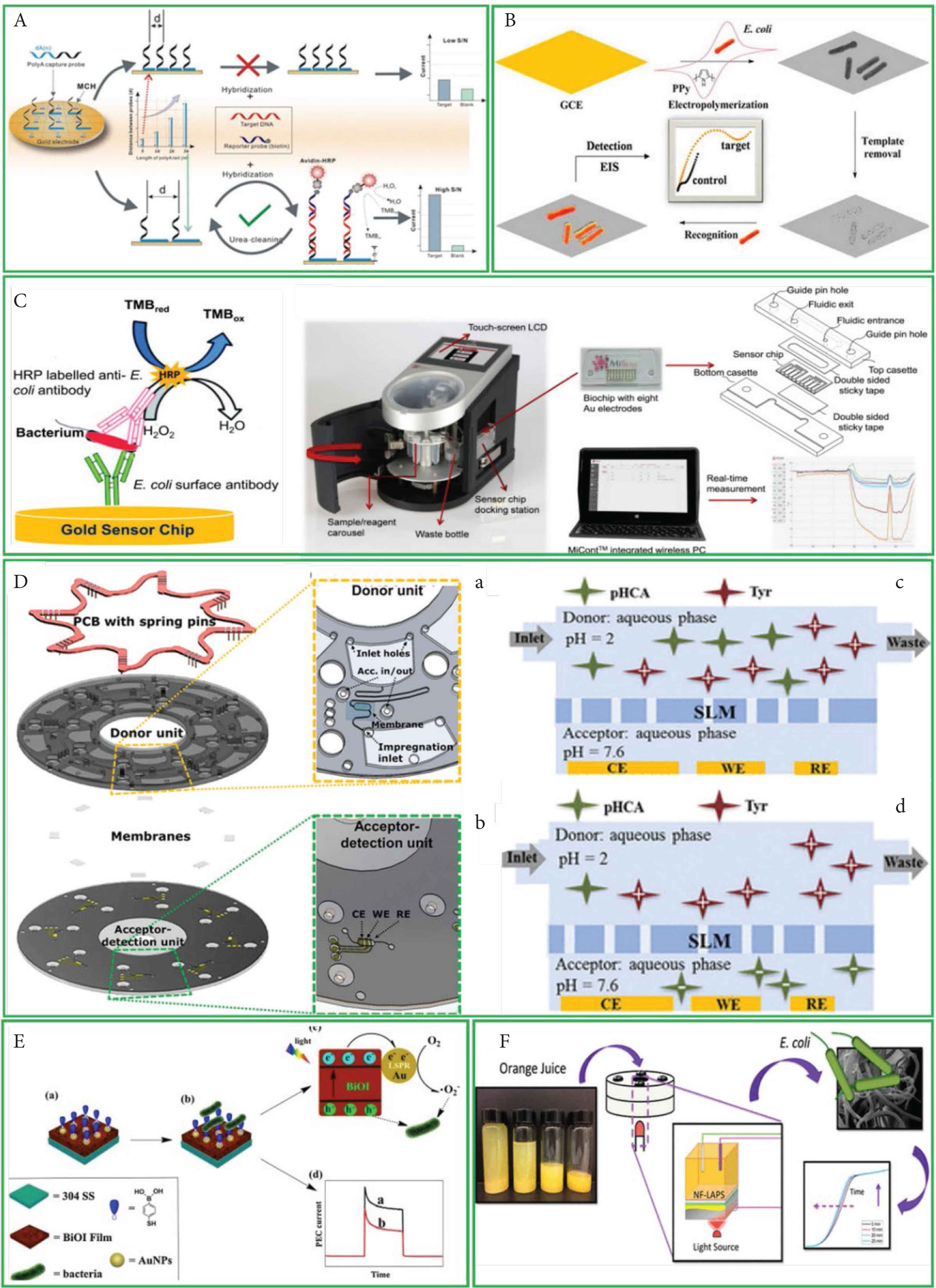
(A) Electrochemical DNA sensor based on the assembling strategy using a poly A capture probe [33]. (B) The construction of BIP film-based sensor involving three consecutive procedures: the BIP film preparation, bacterial recognition and impedimetric detection [34]. (C) Illustration of custom-designed MiSens biosensor integrating microfluidic system, biochip design and real-time amperometry [27]. (D) Inner diagram of the lab-on-a-disc sensor with integrated supported liquid membrane (SLM) extraction and embedded electrodes for detection; a) Illustrative diagram of the donor unit and b) acceptor-detection unit; c) pHCA becomes neutral, while Tyr (existing basic amino group) is positively charged in the acceptor unit; d) During the extraction process, neutral pHCA diffuses to the SLM to the acceptor site, where it becomes negatively charged [28]. (E) Photoelectrochemical system for capture, detection, and inactivation of E. coli [36]. (F) LAPS sensor using a Si-based chip with pH sensitive hydrogel nanofibers [29].
To date, electrochemical microfluidics sensors incorporating sample pretreatment modules and detection modules have achieved fully automated or semi-automated detection of E. coli. For example, a fully automated microfluidic-based amperometry has applied for portable and on-site detection of E. coli in water sample [27]. The use of gold nanoparticles (AuNPs) conjugated antibody significantly enhanced the sensing performance (Figure 1C). The sensitivity of the device to E. coli was found to be 50 CFU/mL in water sample. The sensor could reuse the same sensing surface to test different bacteria concentrations. Furthermore, this study has excellent potential for the quantification of food pathogens and clinical sample analysis. Genetically modified E. coli expressing tyrosine (Tyr) ammonia-lyase could convert Tyr to hydroxycinnamic acid (pHCA). In order to realize efficient detection of pHCA in complex samples, there was a centrifugal fluidic platform consisting of supported liquid membrane extraction and SWV detection (Figure 1D) [28]. Although this method is beneficial for online monitoring of bacterial bioprocess, it can only test single sample in a small volume (3 μL), which requires the modification of nanomaterials on the electrode to improve the sensitivity of the sensing system. Hence, a lab-on-a-disc platform integrated eight sample filtration and electrochemical detection for pHCA [35]. Due to the proximity of the redox potentials of the pHCA and the substrate (Tyr), the filtration device was used to screen pHCA from the culture medium. The platform could availably quantify the concentration of pHCA in the range from 0.125 to 2 mM. Moreover, the sensor is expected to achieve at-time monitoring the dynamic change of pHCA in the biological process.
Photoelectrochemical platforms have attracted extremely interest in sensing analysis owing to its low cost, rapid response, and high sensitivity. Researchers have integrated optical materials into sensors and devoted to improve sensing performance. There were photo-electricity conversion unit, visible light driven (VLD) photocatalytic antibacterial unit, AuNPs link unit and capture unit in a multifunctional photoelectrochemical system (Figure 1E) [36]. E. coli could be captured on the photoelectrode surface by the reversible binding between boronic acid group and peptidoglycan of the bacteria cell wall, which causes a decrease of photocurrent owing to the steric hindrance blocking the transfer of electron donor to photoelectrode surface. This technology has a profound impact on the application of VLD photocatalytic material-based photoelectrochemical sensor. Besides, a portable device integrating hydrogel nanofibers with light addressable potentiometric sensor (LAPS) was developed in food safety application (Figure 1F) [29]. The sensitivity of the sensor toward E. coli in undiluted orange juice was found to be 102 CFU/mL. The designed nanofibers-LAPS has been a promising technology to ensure other fruit juices safety at different processes of production, distribution, and consumption.
3.2. Electrochemical Sensors for Detection of Salmonella
Genus Salmonella is an important member of the family enterobacteriaceae, which consists of Salmonella enterica (S. enterica) and Salmonella bongori [37]. In general, human infections resulting from 2500 Salmonella serovars are particularly in connection with contaminated foods typically pork, eggs, poultry, vegetables, and fresh fruits. Common symptoms, such as typhoid fever, paratyphoid fever and gastroenteritis, have drawn correlative researchers’ attention to food safety problems caused by Salmonella [38].
Silva et al. [39] integrated polymer membrane solid layer enveloping AuNPs-antibody on miniaturized ion selective electrodes (i.e., pipette tip electrode) to fabricate an electrochemical immunosensor. When the antibody interacts with Salmonella typhimurium (S. typhi) to stable state, it blocks selective ions (non-redox activity) mass transfer to the bulk solution, resulting in electromotive force change in aqueous layer (Figure 2A). The designed sensor was used to directly analyze bacteria from commercial apple juice in complete analysis time, less than 1 h. A lower detection limit of 6 cells/mL was obtained. Due to its low cost, label-free strategy and fast response, the sensor can be a good prototype device for responsing different pathogenic bacteria. In addition to the pipe tip electrode, LIG electrode has been successfully applied to the construction of S. typhi immunosensor due to its unique high electrical conductivity, chemical stability, low cost, and rapid synthesis under loose conditions [24]. The immunosensor was attained through laser inducing polyimide film to form porous graphene electrode in ambient conditions and antibody functionalization on LIG electrodes in turn (Figure 2B). LIG biosensor could detect live bacteria from chicken soup in a wide range of 25–105 CFU/mL with a low detection limit of 13 CFU/mL. The results demonstrated that the sensing method was a viable option for ensuring uncontaminated foods reach the consumer.
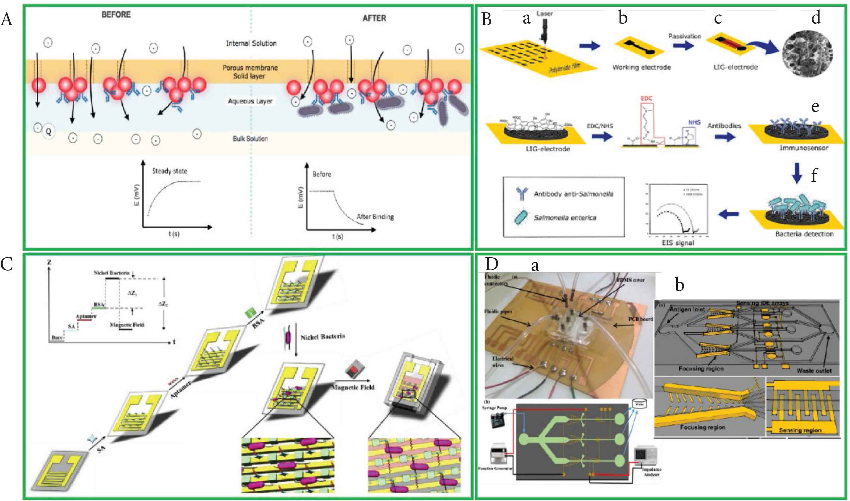
(A) Schematic of surface blocking effect detection mechanism on sensing interface [39]. (B) Fabrication, biofunctionalization, and sensing scheme of the LIG immunosensor [24]. (C) Electrochemical biosensor based on aptamer-bacteria-nickel nanowire complexes on interdigitated microelectrode [41]. (D) a) Completely fabricated bonded biosensor showing the fluidic connectors and tubes; b) Schematic images of impedance-based sensor, magnified view showing the focusing electrode, and magnified view of the detection electrodes [44].
As we know, the combination of pure electrodes and biological receptors could hardly achieve the high sensitivity detection of Salmonella. Therefore, more researches have focused on functionalized nanomaterials, magnetic separation techniques, and amplification reactions [polymerase chain reaction (PCR); helicase-dependent amplification (HDA)] to design signal amplification strategies. Barreda-García et al. [40] fabricated a HDA-electrochemical genosensor to improve Salmonella analysis to the single copy level. The detectability of this sensor improved in two-fold of the real-time PCR. The other sensitive sensor was based on aptamer coated gold interdigitated microelectrode and nickel nanowire binding to antibody for Salmonella capture and separation, respectively (Figure 2C) [41]. The detection limit of 80 CFU/mL was obtained in the range from 102–106 CFU/mL. Besides, Bu et al. [42] encapsulated ferrocenes into AMPs-Cu3(PO4)2 nanocomposites as signal amplification probe. Antibody-coated magnetic beads were used to capture and concentrate the target cells. The result showed a low detection limit of 3 CFU/mL and a linear range from 10–107 CFU/mL.
Microfluidic technology integrating immune separation and enrichment of bacteria could implement simultaneous measurements of multiple analytes, which is expected to realize lab-on-a-chip system for on-site analysis of Salmonella. The constructed hundreds of disposable microfluidic devices were feasible to achieve simultaneous measurement of eight samples with a low LOD of 7.7 cells/mL, in a linear range from 10–100 cells/mL [43]. And other researchers used positive dielectrophoresis to concentrate the antigens for highly sensitive detection of three Salmonella serogroups with a LOD of 7 cells/mL [44]. The sensing principle was the binding of antigen to antibody, leading to the impedance signal change (Figure 2D). This microfluidic sensor could differentiate live bacteria from the dead ones, which was applied in process control in slaughter processing plants.
3.3. Electrochemical Sensors for Detection of Staphylococcus aureus
S. aureus is a representative of gram-positive bacterium and is a common foodborne pathogenic microorganism [45]. Under appropriate conditions, it can produce enterotoxin and cause food poisoning with symptoms of nausea, vomiting, diarrhea, and dehydration. S. aureus is also the main culprit for furunculosis [8]. Moreover, mutations in the gene sequence of S. aureus could lead to the production of drug-resistant strains (methicillin-resistant S. aureus, MRSA), and further resistance against β-lactamases [46]. Therefore, timely prevention and sensitive detection technology is the most effective measurement to deal with the outbreak of S. aureus and drug-resistant strains.
Recently, quantitative analysis of S. aureus has focused on ultrasensitive detection and multifunctional sensing platform. Cai et al. [26] utilized strand displacement amplification and triple-helix molecular switch to determinate S. aureus. Guanine (G)-rich probe was anchored to the stem of molecule switch to form triple-helix DNA structure, which could improve sensitivity and prevent guanine tetramer formation. The strand shift amplification technique was used to release more single stranded DNA (ssDNA) that was completely complementary to the ring region of the molecular switch, resulting in the probe being exposed to the solution to form guanine tetramer. The guanine tetramer could bind to heme to form the electroactive complex (Figure 3A). This sensor provided a high sensitivity and a low detection limit of 8 CFU/mL. And the sensor was applied for S. aureus detection in lake water, tap water and honey samples. Based on the unique charge properties of bacteriophages, Farooq et al. [47] orientated phages onto the electrode surface modified nanocomposites, highly porous bacterial cellulose (BC)/carboxylated multiwalled carbon nanotubes (c-MWCNTs). BC with highly porous and fibrous, offered a huge surface area for the impregnation of c-MWCNTs. Polyethyleneimine (PEI)-functionalized nanocomposites could identify the head of phages with negative electricity. The exposed tail was used to capture bacteria (Figure 3B). The DPV based biosensor could detect 3 CFU/mL and 5 CFU/mL of S. aureus in phosphate buffer saline and milk, respectively. And the biosensor provided a sensitive, specific, and accurate tool for early detection of S. aureus in food samples. Multifunctional analysis platform integrating simultaneous detection, elimination, and inactivation of pathogens was an efficient technology [48]. Vancomycin was specific to peptidoglycan on the bacteria cell wall to capture target cells. Silver ions released by silver nanoparticles (AgNPs) were able to kill bacteria (Figure 3C). The platform relied on the vancomycin-functionalized AgNPs/ZnO nanorod arrays can measure S. aureus with a LOD of 3.3 × 10² CFU/mL. It has profound significance using multifunctional biosensor to implement pathogens analysis in water.
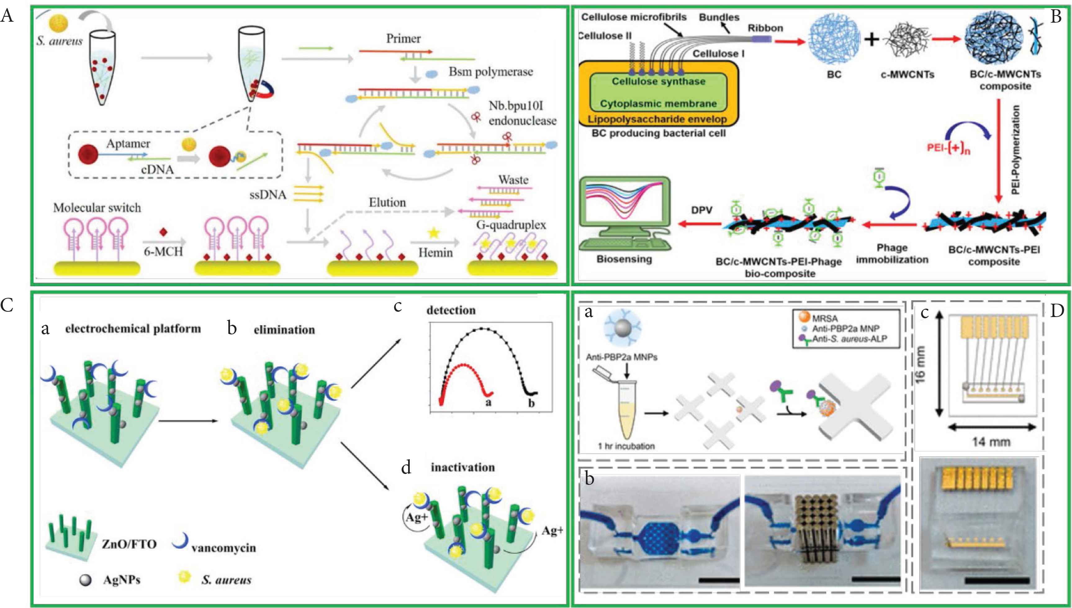
(A) Scheme of the electrochemical biosensor for S. aureus based on triple-helix molecular switch [26]. (B) Design process of ultrasensitive sensor: BC production, incorporation of c-MWCNTs into its matrix, its cationic modification with PEI, immobilization of phages in the PEI-modified BC fibers, and DPV detection [47]. (C) Multifunctional electrochemical platform for simultaneous detection, elimination, and inactivation of S. aureus [48]. (D) a) Schematic representation of the microfluidic platform including bacterial capture unit and electrochemical detection unit (b, c) [30].
Besides, multi-signal probes (MSP) system for determination of mecA genes of MRSA has attracted our attention [46]. In MSP system, the use of seven biotin-labelled signal probes remarkably improved the approachability of target sequence embedded in complex DNA structures. The MSP system could improve stability of the entire system by optimizing the density of capture probes. In comparison with other gene sensors [49,50], this sensor had a lower LOD, 57 fM mecA genes. However, it is only prospect for instrument to achieve microminiaturization and on-site monitoring of real sample. Subsequently, Nemr et al. [30] integrated magnetic capture unit and electrochemical detection unit in a microfluidic platform (Figure 3D). Due to penicillin-binding protein 2a (PBP2a) leading to methicillin resistance, anti-PBP2a antibodies functionalized magnetic nanoparticles was used to specifically capture MRSA. Then, alkaline phosphatase (ALP)-functionalized anti-S. aureus antibodies could recognize magnetic MRSA. This technique has been successfully used in clinical diagnosis and has great potentiality to accommodate different bacteria.
3.4. Electrochemical Sensors for Detection of Listeria monocytogenes
Listeria monocytogenes, opportunistic foodborne pathogens, generally exist in ready-to-eat foods, such as seafood products, milk products, and heat-treated meat products [51]. Elderly, pregnant women, neonates and immunocompromised population are at higher risk in infecting the disease. Lm could be capable of triggering septicemia, meningitis, abortion, stillbirth, and meningoencephalitis endangering human health [52].
Molecular-based electrochemical biosensors mainly consist of molecular amplification process and electrochemical detection of specific DNA or ribonucleic acid (RNA) sequences in pathogens [52]. Recently, an integrated strategy based on double-stranded DNA PCR amplification products and electrochemical immunoluminescence (ECL) was proposed [53]. An inexpensive and disposable paper-based bipolar electrode was demonstrated to Lm DNA analysis (Figure 4A). The maximum signal of the sensor was observed within 10 s. The sensor had a detection limit of 10 copies/μL of genomic DNA. Moreover, the technology demonstrated advance in biological, clinical, and environmental applications. Among isothermal molecular amplification techniques, the sensitivities of LAMP and recombinase polymerase amplification (RPA) assays were higher than PCR [54,55]. For example, aptamers magnetic capture (AMC)-LAMP could achieve the LOD of 5 CFU/mL. The key advantage of the designed AMC-LAMP was the exemption of sophisticated experiment devices and pre-analytical culture enrichment procedure [54]. Another example, RPA-lateral flow dipstick (LFD) technology was regarded as a solution for POC testing. Although the LFD for pathogens analysis just requires simple equipment, it is limited to researching lots of actual samples [55]. In summary, molecular amplification technology could be used to design electrochemical sensor, which is undoubtedly conducive to the realization of ultrasensitive and POC analysis.
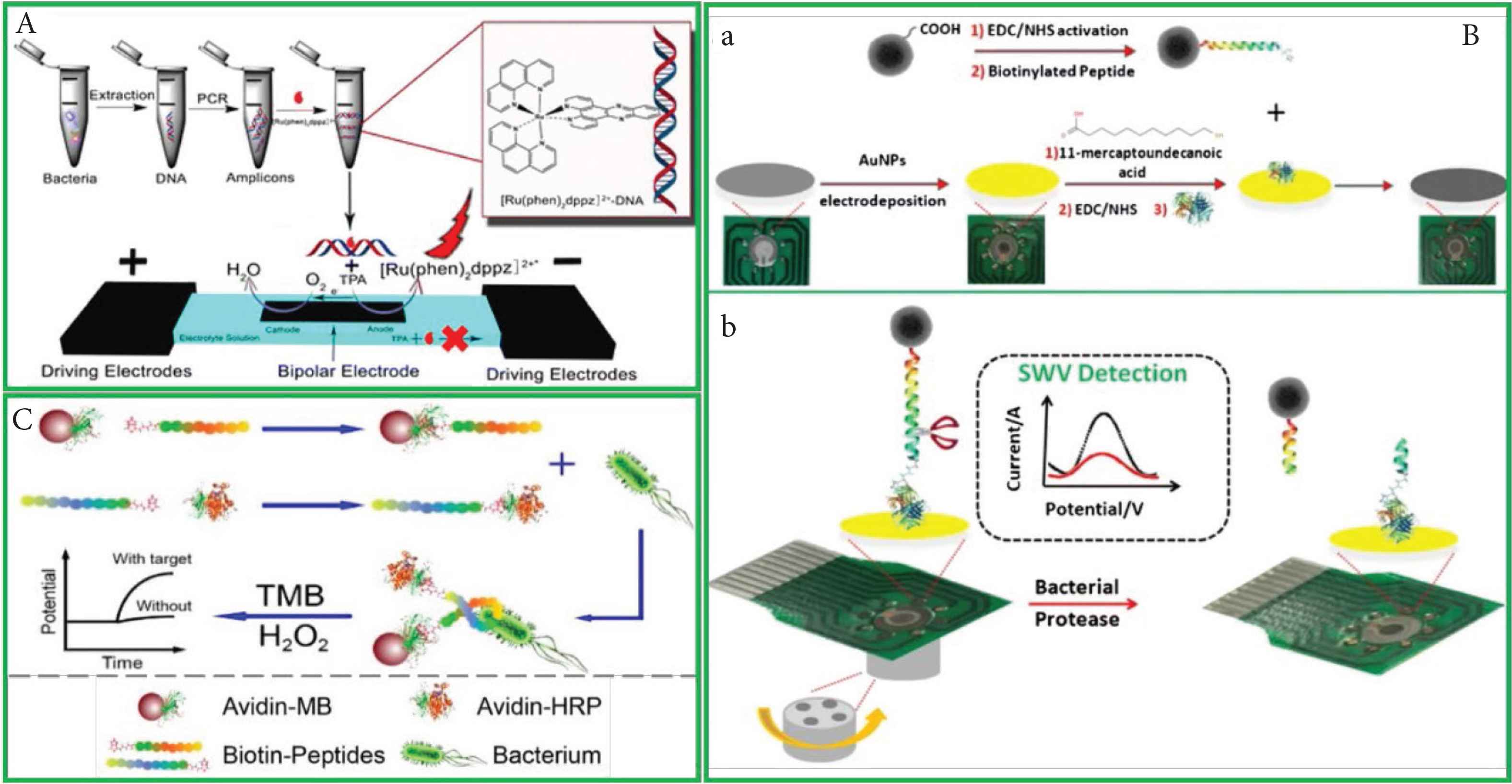
(A) Analysis principle of the bipolar electrode-ECL molecular switch system [53]. (B) Scheme of the multiplexed array biosensor. (a) The fabrication process of peptide-magnetic bead/AuNPs/SPCE. (b) Electrochemical detection mechanism: the specific cleavage of peptides through bacterial proteases causing the changes in electrical signals on the sensor surface [56]. (C) Scheme of the potentiometric sandwich assay based on short antimicrobial peptide pairs [57].
Recently, there was an electrochemical biosensor able to perform multiplexed detection of Lm using peptide as recognition receptor and proteases enzyme produced from bacteria as marker [56]. Each protease enzyme had the capability to break a peptide bond at specific site causing a dissociation of the magnetic nanoparticles from the electrode surface which can be detected by SWV (Figure 4B). This biosensing system enabled to achieve fast and multiplexed analysis for Lm, with a detection limit of 9 CFU/mL. Compared to multiple peptides, short peptides had high specificity for the target. Moreover, the short peptides with less positive charges could also reduce the adsorption of interferences species in real samples (such as other bacteria and negatively charged species). And thus, Lv et al. [57] used short peptide pairs obtained from splitting the original peptides to design potentiometric biosensors for Lm (Figure 4C). Similarly, Lm could be detected in a linear range of 102 to 106 CFU/mL, with a LOD of 10 CFU/mL. This approach broadened the applications of peptides-based pathogen biosensors in the field of food safety.
Polyclonal antibody (PAb) may identify multiple epitopes on any antigen, while a monoclonal antibody (MAb) may detect only one epitope on any antigen [58]. The general principles are as follows: (1) magnetic nanomaterials based on PAb specifically capture target bacteria and isolate them; (2) MAb-based AuNPs carry catalytic materials, such as urease, to catalyze the reaction of electroactive material. A low-cost screen-printed electrode was used to detect Lm in the range of 1.9 × 10³–1.9 × 10⁶ CFU/mL. The designed sensor was suitable for in-field analysis [59]. Additionally, Chiriacò et al. [60] designed a miniaturized biochip integrating an array of interdigitated antibodies-functionalized electrodes for Lm analysis in clinical and on-site detection. The biochip achieved high-throughput and high sensitivity with a LOD of 5.5 CFU/mL. However, it requires data processing skills and simple samples. Therefore, future work should focus on addressing these challenges and obstacles.
3.5. Electrochemical Sensors for Detection of Other Bacteria
Vibrio parahaemolyticus (Vp), one gram-negative halophilic acrogenous bacterium, was found in zooplankton, coastal fish, and shellfish. Once Vp-contaminated seafood absorbed via the body, it would cause severe diarrheal disease gastroenteritis, acute gastroenteritis. The presence of Vp would lead to the outbreak of foodborne diseases and endanger the safety of human life. Teng et al. [61] developed an antibody-aptamer based electrochemical sensor for ultrasensitive detection of Vp. This protocol provided a versatile platform and detected the lower concentration of 2 CFU/mL in spiked fish samples. But the fabrication procedures of sensor still need further simplification. Fortunately, a POC device integrating a diminutive potentiostat and unmodified screen-printed graphene electrodes (SPGE) can effectively detect LAMP amplicons [62]. In addition, ECL immunoassay with high sensitivity has been employed for Vp detection with a detection limit of 5 CFU/g for seafood [63]. However, ECL response is especially sensitive to nonspecific substance, leading to relatively low reliability. Therefore, researchers provided a Faraday cage-type immunosensor to achieve ECL and anodic stripping voltammetry dual-modal detection, which could improve the reliability of sensors [64]. The Faraday cage-type structure could enhance signal effects. The results showed a detection limit of 33 CFU/mL.
Campylobacter jejuni, Campylobacter hepaticus, and Campylobacter coli belong to Campylobacter species, all playing their part in foodborne diseases associated with human. Lately, researchers explored the effects of single leg skin samples and pooled neck skin samples from on the detection of Campylobacter contaminated chicken. The results showed that, by changing from single leg skin to the pooled neck skin, the estimated sensitivity and prevalence could both increase by ~1.6 times [65]. A multiplex PCR was designed to simultaneously identify the presence of different Campylobacter species in chicken samples. The assay may be a new and viable diagnostic tool for evaluating bacteria contaminated meat product [66]. However, there are few papers based on electrochemical biosensor to detect Campylobacter.
Cholera is an acute diarrhoeal infectious disease caused by the ingestion of food or water contaminated with Vibrio cholerae. To satisfy POC diagnosis such as higher levels of sensitivity and specificity, simplification, portability and low cost, Valera et al. [67] reported an on-chip biosensing platform for the detection of cholera toxin subunit B. Gold dendrites functionalized via poly(2-cyanoethyl)pyrrole (PCEPy) was used as working electrode and could allow for a higher level of detection sensitivity. This sensor was more sensitive (detection limit of 1 ng/mL) than detection using a simple planar gold electrode (detection limit of 100 ng/mL). The on-chip device represents a promising avenue for POC disease diagnosis in resource limited areas. Further work is toward realization as an on-chip diagnostic device integrating microfluidic technology.
Besides, researchers designed an aptasensor for low-cost and highly specific response of Shigella dysenteriae in dairy products [68]. Aptamers were assembled on AuNPs modified electrode. Moreover, the fabricated biosensor had an admirable sensitivity with a LOD of 1 CFU/mL. Due to its good performance, it could be a practical tool in food or clinical quality control. Also, Yuan et al. [69] designed a cascade signal amplification based on reverse transcription (RT)-PCR triggering G-quadruplex DNAzyme catalyzed reaction to determine Cronobacter sakazakii. Only tDNA could initiate RT-PCR reaction and the G-quadruplex binding with hemin assembled an artificial DNAzyme to catalyze the oxidation of 3,3′,5,5′-tetramethylbenzidine by H2O2. The electrochemical sensor could detect Cronobacter sakazakii with a detection limit of 5.01 × 10² CFU/mL. Due to the advantages of high sensitivity, low cost and simple manipulation, this approach provides the option of potential application in other pathogen detection.
4. CRITICAL DISCUSSIONS
In the last decade, most of the work was devoted to synthesizing new functional materials and designing novel sensing principle that greatly enhance sensitivity, selectivity, overpotential reduction and stability. Though there is considerable improvement in the applications of electrochemical sensors for foodborne bacteria detection, there are few limitations, which hinder these applications for the next level. The specific limitations are as follows.
Nucleic acids and aptamers are used as bioreceptor elements and retain its long last activity, which is a critical issue in front of the researchers. The sustainability of sensing materials (such as nanomaterials) in sensor applications, has been insufficiently investigated. The fabrication process and toxicity of functional materials change according to the physical properties of the material type [100]. Therefore, improving the stability of bioreceptor elements and functional materials, and then developing the service life of sensors are the critical issue worthy of investigation by researchers.
Highly sensitive detection of foodborne bacteria is still a goal pursued by researchers. Small volume samples, such as skin, intestines, intestinal mucosa and other special occasions of the human body [101,102], could be detected using highly sensitive devices. It is feasible to dilute the actual sample (small volume) and then test it. And the sensor with such lower detection limit is conducive to rapidly screen suspected patients. Of course, high sensitivity may mean poor accuracy, so how to balance the sensitivity and accuracy of the sensing method needs to be considered by researchers in the relevant field. Besides, the nonspecific adsorption of the sensors in the food complex matrix could lead to poor accuracy of detection results. Thus, it is inevitable to carry on tedious pretreatment to the actual sample. Currently, very few sensors have attained commercial success, apart from electrochemical glucose sensors and lateral flow pregnancy tests [100,103]. Electrochemical bacteria sensor integrated into affordable cost, miniaturization, portability, high accuracy and easy operability still remain challenges.
5. CONCLUSION AND FUTURE PROSPECTS
Foodborne pathogenic bacteria continue to hold their viability and express their poisonousness. It is extremely significant for timely prevention, specific identification, sensitive and rapid response (or online monitoring) of pathogens. Electrochemical sensor is a sustainable and indispensable diagnostic tool for pathogens analysis. As an ideal electrochemistry sensor, it needs to meet the following requirements: (1) the interface design of the sensor is preferably to have the advantages of simple operation, low cost, high specificity, and large surface area; (2) the whole system satisfies miniaturization, commercialization, functional integration, full automatization, and wide application. Nowadays, only a few sensors have satisfied this criterion, but most sensors are still facing challenges.
Selectivity is one of the key factors to determine the performance of sensors. At present, AMPs as low-cost biorecognition elements obtained from natural substances (such as plants and microorganisms) are attractive candidates. In addition, short peptides replacing long chain peptides could efficiently improve the specificity of devices. However, current methods of interface design have limitations to the stability and repeatability of analytical results. We can combine contactless 3D printing technology to the sensing interface to improve analytical accuracy, reproducibility, and service life of the sensor. Also, the integration of artificial intelligence will greatly enrich the data module. Significantly, high sensitivity has always been the most prominent characteristic of electrochemical sensing over other analytical methods. Micro-machining technology (like DNA walking machine) combined with new active nanomaterials and optical materials could improve the sensitivity of the system.
It is noted that only several sensors have been commercialized and evolved conceptual approaches into practical applications. Miniaturized equipment for on-site analysis is a luxury for the public. During the whole analysis process, the culture of bacterial strain often requires controlled temperature and specific environment, which is time-consuming in spite of being sensitive and accurate. Of course, most pre-treatment process of real samples cannot detach from the standard laboratory. Future researchers could combine artificial intelligence technology to solve the confusion of practical application. At last, we hope to provide a bridge to connect the gap between food science and electrochemical sensors.
CONFLICTS OF INTEREST
The authors declare they have no conflicts of interest.
AUTHORS’ CONTRIBUTION
YW contributed in investigation, design, review, writing and revision. XM, XQ and PY contributed in investigation and discussion. QS, MZ and TY contributed in conceptualization, supervision, revision, editing and critical review.
ACKNOWLEDGMENTS
This work was supported by National Key R&D Program of China (2019YFC1606703) and the Natural Science Foundation of Shaanxi Province in China (2020JM-429).
Footnotes
REFERENCES
Cite this article
TY  - JOUR
AU  - Yahui Wang
AU  - Xin Ma
AU  - Xiujuan Qiao
AU  - Pingping Yang
AU  - Qinglin Sheng
AU  - Ming Zhou
AU  - Tianli Yue
PY  - 2021
DA  - 2021/06/30
TI  - Perspectives for Recognition and Rapid Detection of Foodborne Pathogenic Bacteria Based on Electrochemical Sensors
JO  - eFood
SP  - 125
EP  - 139
VL  - 2
IS  - 3
SN  - 2666-3066
UR  - https://doi.org/10.2991/efood.k.210621.001
DO  - 10.2991/efood.k.210621.001
ID  - Wang2021
ER  -