
A clinical study of cutaneous changes in pregnancy
- DOI
- 10.1016/j.jegh.2016.10.002How to use a DOI?
- Keywords
- Pregnancy dermatoses; Specific dermatoses
- Abstract
Background/objective: Pregnant women experience a myriad of physiological and metabolic changes that affect different organ systems in the body. Cutaneous and appendageal alterations that manifest during pregnancy are largely modulated by hormonal, immunologic, and metabolic factors. Detailed reports encompassing physiological changes and specific dermatoses of pregnancy and effects of various dermatoses on pregnant women are scanty in literature. This study was conducted to examine in detail both physiological changes and specific dermatoses. The cutaneous changes are divided into physiological changes, skin diseases aggravated by pregnancy, and specific dermatoses of pregnancy. The objectives were to study the various cutaneous changes of pregnancy and to know the proportion of these cutaneous manifestations in pregnant women.
Methods: This study included 600 pregnant women attending the Obstetrics and Gynecology Department of a tertiary teaching hospital in Northern Kerala, India. Detailed history elicitation and complete physical and dermatological examination were performed. Skin biopsy was performed in relevant cases.
Results: Cutaneous changes were seen in a majority of patients, of which physiological changes were the most common (99%). The most common cutaneous manifestation was hyperpigmentation (526; 87.6%), followed by striae gravidarum (72.8%). Other changes were vascular, including pedal edema (10%), pregnancy gingivitis (1.8%), and varicose veins (1%). Infections were the common dermatological problem in this study group. The most common infections were vulvovaginal candidiasis (21%), Tinea versicolor (6%), scabies (2.8%), dermatophytosis (1.5%), and sexually transmitted infection (0.5%). Specific dermatoses were seen in 12 cases (2%), with the most common being pruritic urticarial papules and plaques of pregnancy (1.3%).
Conclusion: Pregnant women are prone to suffer from a wide range of dermatological problems apart from specific dermatoses of pregnancy. The study emphasizes the need for a detailed and meticulous examination of these patients to detect these various disorders.
- Copyright
- © 2016 Ministry of Health, Saudi Arabia. Published by Elsevier Ltd.
- Open Access
- This is an open access article under the CC BY-NC-ND license (http://creativecommons.org/licenses/by-nc-nd/4.0/).
1. Introduction
Pregnancy is accompanied by profound immunologic, metabolic, endocrine, and vascular changes, which make pregnant women susceptible to changes of skin and appendages, both physiologic and pathologic. These changes are a positive adaptation of the mother to accommodate and support the fetus as it grows and develops throughout gestation [1]. Many studies have focused on a particular dermatosis or other diseases and conditions related to pregnancy [2–6]. However, detailed reports encompassing the physiological changes and specific dermatoses of pregnancy and effects of various dermatoses on these women are scanty in literature. Existing studies show a wide range of variations in the incidence of specific dermatoses of pregnancy. For these reasons, a clinical study was conducted to study both physiological changes and specific dermatoses of pregnancy.
2. Materials and methods
An observational cross-sectional study was conducted, which included 600 consecutive pregnant women attending the Obstetrics and Gynecology Department of Government Medical College, Calicut, Kerala. Ethics Committee clearance and informed consent of patients were obtained. Detailed medical history of patients including chief complaints related to presence of pruritus, onset of skin lesions in relation to duration of pregnancy, jaundice, vaginal discharge, and associated medical or skin conditions was noted. Obstetric details such as duration of pregnancy and obstetric score were also noted. Detailed and complete physical examination of all patients was performed. In addition, a detailed dermatological examination was performed for patients (by V.V.P.) to look for physiological changes and specific dermatoses. If any specific dermatoses were present, the morphology of skin lesions, distribution, and sites involved were studied. The diagnosis was based mainly on clinical grounds; skin biopsy was done for confirmation in doubtful cases only. If the patient gave a history of a pre-existing skin disease, any evidence of exacerbation or remission was noted, and relevant systemic examination was performed. The recruited patients were re-examined if they presented with any fresh complaints. A routine examination of blood and urine and screening for human immunodeficiency virus and venereal disease were done in all cases. Other necessary investigations such as fungal scraping, KOH examination of vaginal discharge, Tzanck test, and skin biopsy were done whenever necessary.
3. Results
A total of 600 patients were included in this study. The age of our study population ranged from 18 years to 38 years, with a majority being in the 20–25-year age group. Most cases were primigravida (370, 61.67%); 230 (38.33%) were multigravida. Most cases presented in the second trimester (400, 66.67%). A majority had no skin-related complaints. The most common complaint encountered was pruritus (113, 18.83%), followed by presence of skin lesions (48, 8%). Other complaints included vaginal discharge and genital lesions. Among those who had pruritus, the commonest cause was fungal infections in 63 cases (55.7%); other causes were scabies (17, 15%), specific dermatoses (10.6%), and miliaria (10, 8.8%). No causes could be identified in 9.7% of the cases.
Pregnancy dermatoses were divided into three categories, namely, physiological changes, skin diseases affected by pregnancy, and specific dermatoses of pregnancy.
3.1. Physiological changes
Physiological changes (Table 1) were seen in a majority of the cases (594, 99%). Those who did not have any changes were primigravida in the first trimester of pregnancy. Among the various physiological changes, the most common manifestation was hyperpigmentation, which was seen in 526 (87.67%) of the cases (Table 1). Of these, linea nigra (Fig. 1) was the most common pattern, noted in 526 cases (87.67%). Other changes seen were secondary areola (235; 39%; Fig. 2), pigmentation of neck (186; 31%), flexures and genitalia (168; 28%), and melasma (13; 2.16%). Of the 526 women with pigmentation (Fig. 3), most were in the third trimester. Pigment changes were seen in 325 (87.8%) and 201 (88%) multigravidas and primigravidas, respectively. There were 437 cases with striae distensae; 256 (58.88%) had white atrophic striae (Fig. 4) and 181 (41.41%) had purplish striae (Fig. 5). The most common site of these changes in all cases was the lower abdomen. In cases with purplish striae, 163 were in the third trimester and 18 were in the second trimester, with none in the first trimester. Atrophic white striae showed almost a similar frequency in all the three trimesters. In primigravidas, purplish striae was common (45.67%), whereas in multigravidas, atrophic striae was common (76.95%). Vascular changes seen included bilateral pitting pedal edema (63, 10.5%), pregnancy gingivitis, and varicose veins. Glandular changes seen were Montgomery’s tubercles (56, 9.33%) and miliaria (8, 1.3%). Of the 600 women, most did not have any history of hair changes; 37 (6.16%) women, however, reported a history of hair changes. Of these, 26 (4.8%) patients noticed improvement in scalp hair, whereas eight (1.2%) reported increased hair loss.
| Physiological changes | No. of patients | % |
|---|---|---|
| Hyperpigmentation | 526/600 | 87.67 |
| Striae distensae | 437/600 | 72.83 |
| Vascular changes | 80/600 | 13.33 |
| Glandular changes | 61/600 | 10.16 |
Physiological changes.
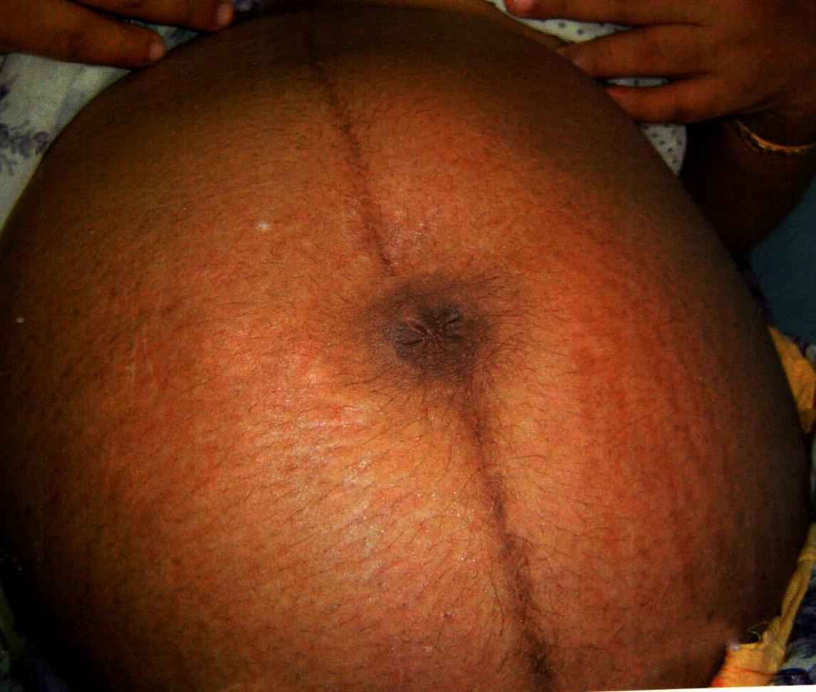
Linea nigra.

Secondary areola.

Flexural pigmentation.

Whitish striae.

Purplish striae.
3.2. Diseases affected by pregnancy
Infections were the most common dermatological problem in our patient group (Table 2). Among the various infections, vulvovaginal candidiasis was the most common (126, 21%). Other infections noted were scabies and dermatophytosis. The sexually transmitted diseases seen were herpes genitalis (0.3%) and condyloma acuminatum (0.16%). One case of systemic lupus erythematosus was seen, which presented with cutaneous flare, but it did not have any adverse effect on the fetus. Acne vulgaris was seen in 62 (10.33%) cases. Most of these patients had exacerbation of acne by the third trimester. Six cases of psoriasis were seen, with the majority being palmoplantar psoriasis. There was one patient with impetigo herpetiformis (Fig. 6), who was a second gravida with an onset of the disease at 32 weeks of gestation.
| Diseases | No. of patients | No. of new patients | No. of patients with pre-existing dermatoses | No. of patients with worsening conditions |
|---|---|---|---|---|
| Infections | ||||
| Candidiasis | 126 | 40 | 86 | 30 |
| Tinea versicolor | 37 | 12 | 25 | 10 |
| Scabies | 17 | 5 | 12 | |
| Molluscum contagiosum | 2 | 2 | ||
| Herpes zoster | 1 | 1 | ||
| Herpes genitalis | 2 | 2 | ||
| Condyloma acuminatum | 1 | 1 | 1 | |
| Autoimmune | ||||
| Systemic lupus erythematosus | 1 | 1 | ||
| Miscellaneous | ||||
| Acne | 62 | 40 | 22 | 20 |
| Skin tag | 52 | 42 | 10 | |
| Seborrheic dermatitis | 7 | 1 | 6 | |
| Neurodermatitis | 3 | 3 | ||
| Pityriasis rosea | 2 | 2 | ||
| Keloid | 1 | 1 | ||
| Psoriasis | 6 | 2 | 4 | 1 |
| Lichen amyloidosis | 1 | 1 | ||
Diseases affected by pregnancy.
Impetigo herpetiformis.
3.3. Specific dermatoses of pregnancy
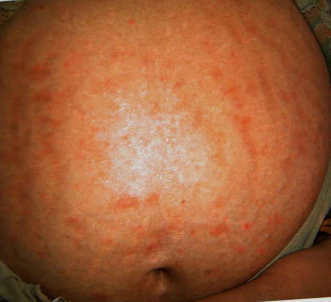
Specific dermatoses were seen in 12 (22%) cases (Table 3). Of these cases, the most common was pruritic urticarial papules and plaques of pregnancy (PUPPP; Fig. 7), which were seen in eight cases (1.3%). All these cases presented between 30 weeks and 36 weeks of gestation, except one case of PUPPP, which presented in the second trimester. Of the eight PUPPP cases, four were multigravida and four were primigravida. The average duration of illness varied from 3 weeks to 4 weeks. There was no adverse effect on the fetus in any of these cases. Other diseases seen were prurigo gestationis (0.3%) and pruritic folliculitis (0.16%); both had onset in the third trimester. The diagnosis of pruritic folliculitis was confirmed by a negative Gram stain and bacterial culture. Of the eight women with PUPPP, five gave a history of atopy. There were three cases of prurigo gestationis (Fig. 8) and all of them had a history of atopy.
| Specific dermatoses | No. of patients | % |
|---|---|---|
| Polymorphic eruption | 8 | 1.3 |
| Prurigo gestationis | 3 | 0.5 |
| Pruritic folliculitis | 1 | 0.16 |
Specific dermatoses of pregnancy.

Pruritic urticarial papules and plaques of pregnancy.
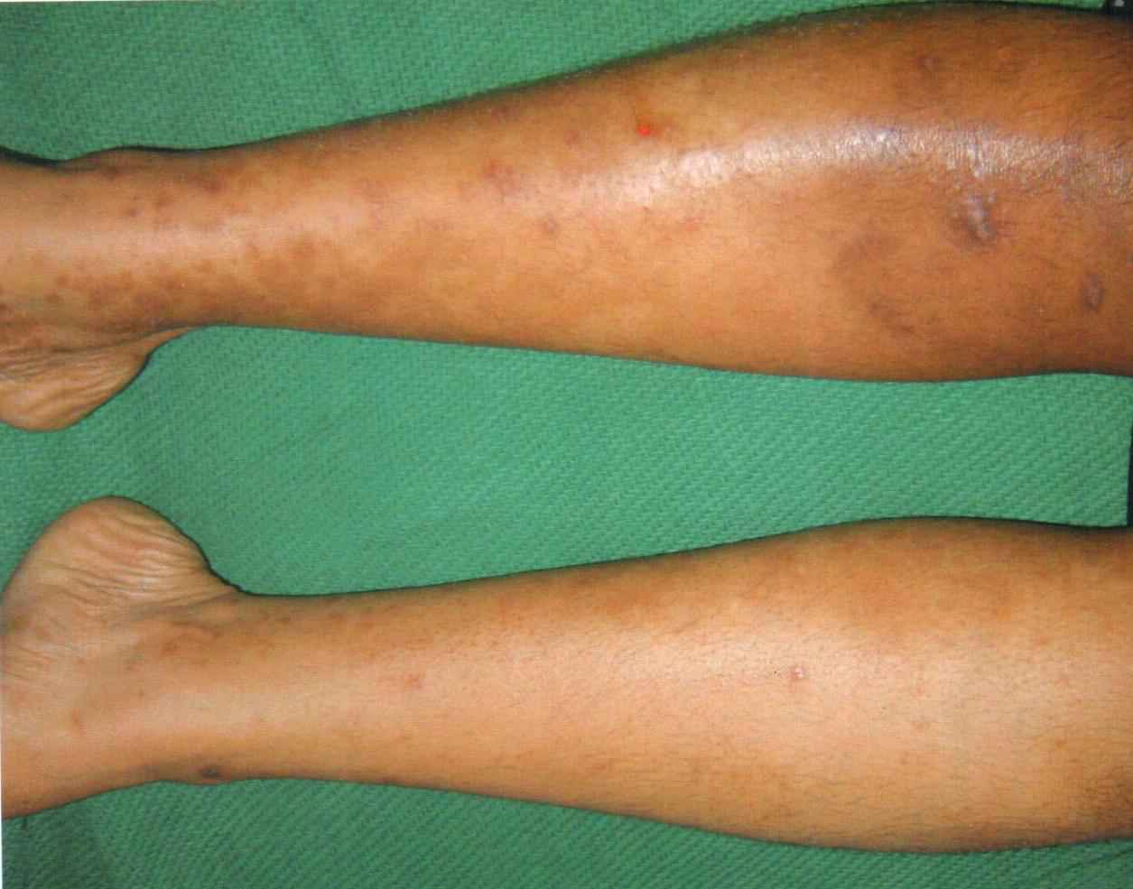
Prurigo gestationis.
4. Discussion
Pregnancy is a unique physiological state characterized by metabolic, immunologic, and hormonal readjustments. These make a pregnant woman vulnerable to all dermatoses occurring in the nonpregnant state and also to certain eruptions related to the physiologic burden of gestation. Apart from these are those skin changes that most pregnant women exhibit, which are considered physiological.
The most common physiological changes are pigment alterations, stretch marks, vascular changes, and telogen effluvium [7]. In this study, most women (594, 99%) experienced physiological changes. In a study by Kumari et al. [8], physiological changes were observed in all patients studied (100%). In the study by Raj et al. [9], which included 1175 pregnant women, only 114 (9.7%) experienced some skin changes. Among the physiological changes, the most common was hyperpigmentation seen in 526 (87.67%) cases. The most common pattern was linea nigra seen in 87.67% of the patients. Even in the study by Kumari et al. [8], the most common pattern of hyperpigmentation was reported to be linea nigra, which was noted in 91.4% of the patients. In our study, pigment changes were seen in 65.27% of the cases during the second trimester and in 99.5% of the cases during the third trimester. The increase in the incidence during the second and third trimesters is because the placenta—the source of estrogen and progesterone, which are strong melanogenic stimulants-starts to function only after 8 weeks of gestation.
Melasma was found in 2.16% of the patients in our study. Winton and Lewis [10] reported the prevalence of melasma in pregnancy to be 15.8%. A malar pattern was seen in 65.9% of cases, whereas a centrofacial pattern was seen in 33.8% of the patients in their study. No case with a mandibular pattern was found. Winton and Lewis [10] reported melasma in 50–75% of pregnant women. The onset of melasma in 12 (80%) cases was in the first trimester in their study, whereas in a study by Martin and Leal-Khouri [7], the onset of melasma was mostly during the second trimester. Wong and Ellis [11] reported melasma in 50–70% of pregnant women with an onset during the second trimester. Muzzaffar et al. [12] reported melasma in 65 (46.4%) patients. However, Indian studies have reported lower incidence of melasma. Raj et al. [9] observed melasma in 10/1175 (8.5%) cases and Kumari et al. [8] reported an incidence of 2.5%. This difference may be because pigmentary changes are more discernible in fair-skinned individuals.
The frequency of striae gravidarum was 72.83% in our study. The atrophic form of striae was common among multigravidas (76.95%), whereas the purplish form was common among primigravidas (45.67%). The onset of purplish striae was during the second trimester with no cases reported in the first trimester. The incidence of atrophic striae was the same in all the three trimesters. Shivakumar and Madhavamurthy [13] reported a frequency of 66.47% for atrophic striae. Kumari et al. [8] reported that striae were seen in 484 (79.7%) cases, of which 217 (44.8%) were primigravidas and 267 (55.2%) were multigravidas.
Vascular changes are congruent with pregnancy because the gravid state increases blood volume, vascular dilatation, capillary permeability, and neovascularization, a process believed to be related to the increase in estrogen and angiogenic factors. In a previous study, nonpitting edema of the legs, eyelids, face, and hands was present in about 50% of women during the third trimester [1]. According to Martin and Leal-Khouri [7], the edema decreases during the day and is thought to be due to secondary sodium and water retention in conjunction with increased capillary permeability. Vascular changes seen in our study include nonpitting edema of feet in 10.5% of the cases and venous varicosities in 1% of the cases. Vascular spiders (spider angiomas) have been reported in 67% of white and 11% of black women between the 2nd and 5th months of pregnancy. Palmar erythema occurs in 66% of white and 33% of black women beginning in first trimester [7]. No cases of palmar erythema were seen in our study. Raj et al. [9] reported the prevalence of palmar erythema to be 33.3%. Bean et al. [14] reported the prevalence to be 62.5% in pregnant white women and 35% in black women. This may be due to erythema being readily appreciated in the light skinned women. A similar discrepancy was found in the observation of spider naevi. The gingival change seen during pregnancy is edema of gums, changing from dark red to blue. Edema and hyperemia may due to hormonal changes as well as local irritation; in some cases, nutritional deficiencies may also be responsible [7]. Pregnancy gingivitis was seen in 11/600 patients in our study. In a study by Muzzaffar et al. [12], 23/140 (16.4%) had gingival edema and redness. Raj et al. [9] reported three cases of pyogenic granulomas. Increased appearance of Montgomery’s tubercles is well-known during pregnancy in 30–50% of pregnant women [7]. In our study, Montgomery’s tubercles were seen in 9.33% of the patients.
We also noted hair changes in 47 women (6.16%). Of these, 29 (4.8%) noticed improvement and lengthening of scalp hair, and eight (1.2%) reported increased hair loss. In the study by Kumari et al. [8], which included 607 women, 11 gave a history of increased hair loss and only five patients noticed lengthening and improvement of their scalp hair, whereas 591 (97.4%) gave history of no change in hair density.
An increased frequency of infection was seen in our study, which is common during pregnancy and is probably related to low cellular immunity. During pregnancy, the cells of the hyperplastic vaginal epithelium get filled with glycogen, desquamate, and contribute to low vaginal acidity, thereby creating an environment suitable for growth of Candida. We found vulvovaginal candidiasis in 21% of patients, which was similar to the findings reported by Shivakumar and Madhavamurthy [13]. Other infections seen were dermatophytosis (21.6%), scabies (2.8%), pityriasis versicolor (6.16%), and molluscum contagiosum (0.3%). Increased eccrine sweating, warm climate, and depressed cellular immunity may be the reasons for higher incidence of these infections [15].
There were three cases of sexually transmitted diseases in our study (2 herpes genitalis and 1 condyloma). The patient with condyloma acuminata was florid due to increased vascularity and moist environment of the vagina.
There was one patient with systemic lupus erythematosus who presented with cutaneous flare. We also observed six (1%) cases of psoriasis. A majority of the cases were palmoplantar pustulosis (4, 0.66%). There was no change in the activity of the disease during pregnancy. There was one case of impetigo herpetiformis. This patient was a second gravida with the onset of disease at 32 weeks of pregnancy. Seborrheic dermatitis was seen in 1.1% of the patients and acne vulgaris in 1.3%; interestingly, there was no change in the activity of these diseases during pregnancy. The increased activity of sebaceous glands could be the cause for these diseases.
Specific dermatoses of pregnancy represent a heterogeneous group of ill-defined pruritic skin diseases unique to pregnancy. Holmes and Black [16] proposed a simplified clinical classification of the specific dermatoses of pregnancy. This classification basically subdivided the specific dermatoses of pregnancy into the following four groups: (1) pemphigoid (herpes) gestationis; (2) polymorphic eruption of pregnancy; (3) prurigo of pregnancy; and (4) pruritic folliculitis of pregnancy [16]. Based on the study conducted by Ambros-Rudolph et al. [17] on 505 pregnant patients, a new classification has been proposed. They have introduced a new term called “atopic eruption of pregnancy,” which covers all patients formerly diagnosed as having prurigo of pregnancy, pruritic folliculitis, and eczema of pregnancy. The incidence of these specific disorders of pregnancy is 0.5–3.0% [18]. In our study of 600 women, 12 (2%) cases of specific dermatoses were seen. Of these, the most common was polymorphic eruption of pregnancy in eight cases (1.3%). In the study by Shivakumar and Madhavamurthy [13], 16 (9.4%) patients had prurigo of pregnancy, six (3.52%) had pruritus gravidarum, and four (2.35%) had polymorphic eruption. In the study by Kumari et al. [8], the incidence of specific dermatoses was 3.6%. In the study by Ambros-Rudolph et al. [17] on 505 patients, various dermatoses were seen, which included eczema of pregnancy (49.7%), polymorphic eruption of pregnancy (21.6%), herpes gestationis (4.2%), pruritic folliculitis (0.2%), and prurigo of pregnancy (0.8%) [17]. The onset of the disease in our study was in the third trimester, except in one case with disease onset at 26 weeks. The average duration of illness was 3–4 weeks. The cases showed good response to treatment with topical steroids and antihistamines and there was no adverse effect on the fetus in any of our cases. Vaughan Jones et al. [19] studied 200 women with dermatoses of pregnancy. In all the cases studied, there was no adverse effect on fetus or mother as a result of pregnancy dermatoses. In a recent Indian study by Puri and Puri [20], the commonest pregnancy-related dermatoses were polymorphic eruption of pregnancy (22%), prurigo of pregnancy (7%), pemphigoid gestationis (3%), pruritic folliculitis of pregnancy (2%), and intrahepatic cholestasis (1%). Another Indian study, however, reported the common pregnancy-specific dermatoses to be prurigo of pregnancy [21].
5. Conclusions
Pregnant women are prone to suffer from a wide range of dermatological problems apart from specific dermatoses of pregnancy. Our study result emphasizes the need for a detailed and meticulous examination of pregnant women to detect these various disorders.
Conflicts of interest
The authors have no conflicts of interest to declare.
References
Cite this article
TY  - JOUR
AU  - Vinitha V. Panicker
AU  - Najeeba Riyaz
AU  - P.K. Balachandran
PY  - 2016
DA  - 2016/11/19
TI  - A clinical study of cutaneous changes in pregnancy
JO  - Journal of Epidemiology and Global Health
SP  - 63
EP  - 70
VL  - 7
IS  - 1
SN  - 2210-6014
UR  - https://doi.org/10.1016/j.jegh.2016.10.002
DO  - 10.1016/j.jegh.2016.10.002
ID  - Panicker2016
ER  -