
Case reports of hydatid disease
- DOI
- 10.1016/j.jegh.2013.01.001How to use a DOI?
- Keywords
- Hydatid cyst; right inguinal region; liver; Echinococcus granulosus
- Abstract
Two cases of hydatid cyst were reported in the Microbiology department, Siddartha Medical College in 2011. 42 year old female presented with a history of painless swelling in the right inguinal region for 5 years which was insidious in onset and gradually increasing in size. She developed itching prior to the hospital visit. 40 year old male presented with abdominal swelling since 1 year, not associated with vomiting or jaundice. Both the patients were from rural background.
Conclusion. Echinococcus granulosus can cause cystic lesions anywhere in the body. High degree of suspicion of hydatid disease is essential in endemic areas in cystic lesions.
- Copyright
- © 2013 Ministry of Health, Saudi Arabia. Published by Elsevier Ltd.
- Open Access
- This is an open access article under the CC BY-NC-ND license (http://creativecommons.org/licenses/by-nc-nd/4.0/).
1. Introduction
Genus Echinococcus has four species that can cause infection in man. These are: E. granulosus, E. multilocularis, E. vogeri and E. oligarthus. E. granulosus is the most widespread and cosmopolitan in distribution. The highest incidence is reported mainly from the sheep-rearing countries. Though India is not primarily a sheep-rearing country, a large number of cases have been reported from Andhra Pradesh and other states of India [1,4,6–11]. Cystic echinococcosis is a zoonosis caused by larval stage of E. granulosus. Man is an accidental host and does not play a role in the biological cycle of the worm. The mode of infection is by the ingestion of food contaminated with dog feces and also by direct contact with dogs. The most common sites affected are the liver (63%), the lungs (25%), followed by muscles (5%), bones (3%), kidney (2%), spleen (1%), and other sites (1%) [2] (Table 1). Infection is usually acquired in childhood. Most cases are asymptomatic. The ingestion by humans of such contaminated food leads to hatching of the ova in the gastrointestinal tract. The enclosed embryos are liberated in the duodenum and transported to the liver by portal circulation. The liver acts as the first filter in trapping the embryos which then develop into hydatid cysts in 55–70% of cases, followed by the lungs as the second filter in 18–35% of cases. Some escape from these filters and develop in other organs [3]. The incubation period is highly variable. The cyst grows at a rate of 0.3–1 cm per year and may take 5–20 years to attain sufficient size to cause symptoms Table 2.
| Organs/tissues | Incidence (%) |
|---|---|
| Liver | 63 |
| Lungs | 25 |
| Muscles | 5 |
| Bones | 3 |
| Kidney | 2 |
| Spleen | 1 |
| Brain | 1 |
| Other sites (Orbit, Breast, Thyroid, Urinary bladder) | 1 |
Incidence of Hydatid disease affecting various organs/tissues.
| S. No. | Case report | Year | Site of lesion |
|---|---|---|---|
| 1 | Arora et al. [6] | 2006 | Muscle |
| 2 | Babu et al. [7] | 2008 | Intraabdominal |
| 3 | Mohan Rao et al. [8] | 2011 | Anterior abdominal wall |
| 4 | Shanthi et al. [9] | 2011 | Spleen |
| 5 | Malik et al. [10] | 2011 | Spleen |
| 6 | Rao et al. [11] | 2012 | Spectrum of hydatid disease in central India |
Review of the literature of Hydatid disease in India.
Case report 1: A rare case of hydatid disease. A 42-year-old female was referred to the Government General Hospital, Vijayawada, Andhra Pradesh from a rural health center with a history of painless swelling in the right inguinal region for 5 years. The onset was insidious and gradually progressive. She developed itching for which she contacted the medical officer of the rural health center. She was advised to undergo ultrasound scanning of the swelling which was suggestive of hydatid cyst of soft tissue. The size was 10 cm in diameter. Routine investigations of total leukocyte count (TLC) and hemoglobin (Hb) percentage were performed; 6700/μl (eosinophils 4%) and Hb% 9.2 mg/ml. Enzyme-Linked Immunosorbent Assay (ELISA), Countercurrent Immunoelectrophoresis (CIEP), Coagglutination, Latex agglutination, and Computerized Tomography (CT) were not done owing to non-availability and financial constraints. Albendazole, the drug of choice was given preoperatively to reduce the risk of recurrence. A cyst arising over the external oblique aponeurosis in the right inguinal region was excised (Figure 1). The contents were sent to the departments of Microbiology (for microscopy) and Pathology (for histopathological examination). A Lactophenol Cotton Blue (LPCB) wet mount and an acid-fast staining were done from a centrifuged [1] deposit of hydatid fluid to demonstrate protoscolices and hooklets (Figure 2) and histopathological reports confirmed the diagnosis (Figures 3 and 4). Post–operatively, the patient was put on Tab. Albendazole 400 mg twice a day for 3 months, and the follow-up has been uneventful.
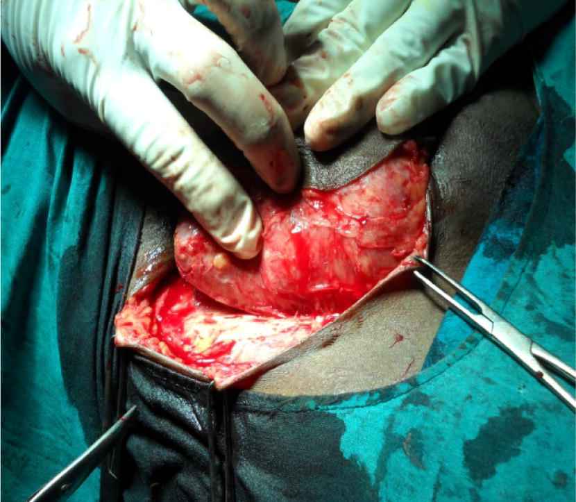
Intraoperative picture of a hydatid cyst.
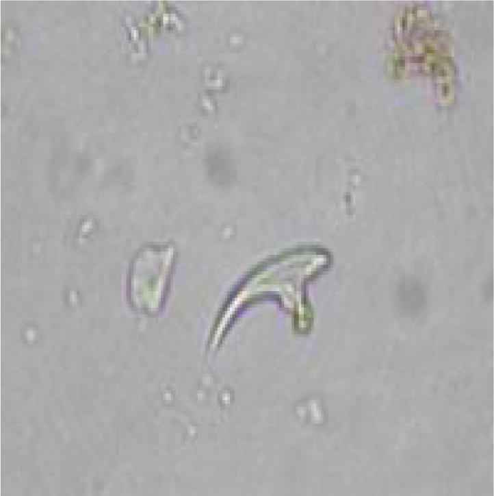
Wet mount demonstrating hooklets in the right inguinal region.
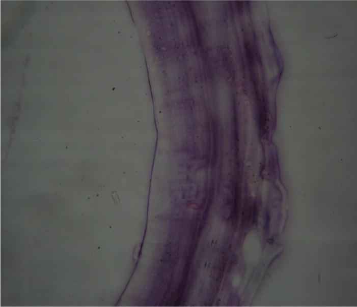
Laminated layer of the ectocyst.

Histopathological section demonstrating hooklets.
Case report 2: A 40-year-old male presented with abdominal pain for 1 year not associated with vomiting and jaundice. No history of fever or weight loss. Abdominal ultrasound suggested cystic swelling in the liver. Routine investigations were completed. TLC 8000/cuμl and eosinophils were 6%. Other investigations were not done owing to financial constraints. He was put on Tab. Albendazole preoperatively to reduce the size. The cyst was excised and sent to the Microbiology and Pathology departments for investigations. The reports – Lactophenol Cotton Blue mount and histopathology – confirmed the diagnosis (Figure 5). The patient recovered from treatment with Tab. Albendazole 400 mg was given twice a day for 4 months.

Cysts and daughter cysts after excision.
2. Discussion
Cystic echinococcosis is still a major problem, especially in rural areas. The condition is mostly asymptomatic. Even though mortality due to echinococcosis is very low, it can produce a very disabling illness. A mortality rate between 0.29% and 0.6% has been reported. In symptomatic cases, the clinical manifestations are highly variable and depend on the following: (a) the organ involved; (b) size and site of the cyst; (c) interactions between expanding cysts and adjacent organs; and (d) complications caused by rupture of the cyst [1,5,6]. Pre-operative diagnosis of cystic echinococcosis is mandatory to prevent anaphylaxis or local recurrence. Ultra Sonogram should be the first imaging choice in abdominal hydatid cysts with sensitivity rates between 93% and 97%. Computerized Tomography should be performed in cases of uncommon locations of the disease. Routine tests like Total Leukocyte count and Hemoglobin percentage should also be done. Moderate eosinophilia 6% or more is usually present. Treatment is essentially surgical. Though liver cysts are common, hydatid cysts in the external aponeurosis are extremely rare. An extremely rare case of hydatid cyst was diagnosed in this area for the first time. However, a number of cases of extra hepatic hydatid disease have been reported from various parts of India, including Andhra Pradesh [6–11] (Figure 6).
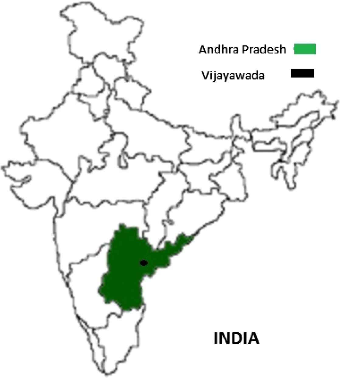
A map of where the cases have been reported.
3. Conclusion
E. granulosus can cause cystic lesions anywhere in the body. Thus, cystic echinococcosis has to be thought of as a differential diagnosis in patients presenting with cystic swellings anywhere in the body in endemic areas unless otherwise proved. Appropriate investigations have to be performed in order to arrive at an accurate diagnosis and in order to prescribe a specific treatment, which is essentially surgical.
Acknowledgment
We are very much grateful to the departments of Surgery and Pathology for their valuable support.
References
Cite this article
TY  - JOUR
AU  - A. Usharani
AU  - G. Deepica
AU  - S. Aruna
AU  - Srinivarao kulkarni
AU  - G. Sai Kamal Kumar
AU  - P. Balamuralikrishna
PY  - 2013
DA  - 2013/03/06
TI  - Case reports of hydatid disease
JO  - Journal of Epidemiology and Global Health
SP  - 63
EP  - 66
VL  - 3
IS  - 2
SN  - 2210-6014
UR  - https://doi.org/10.1016/j.jegh.2013.01.001
DO  - 10.1016/j.jegh.2013.01.001
ID  - Usharani2013
ER  -