
Giant Molluscum contagiosum – A clue to the diagnosis of Human Immunodeficiency Virus infection
- DOI
- 10.1016/j.jegh.2013.06.002How to use a DOI?
- Keywords
- Child; Human immunodeficiency virus infection; Molluscum contagiosum
- Abstract
Molluscum contagiosum (MC) is a benign cutaneous viral infection, affecting mainly children and young adults. Though the disease is self-limiting in immunocompetent individuals, a severe and prolonged course is associated with Human Immunodeficiency Virus (HIV) infection. The following reports an apparently healthy 2-year-old boy with extensive MC without any systemic illness. His parents died of tuberculosis. Investigations revealed him to be a case of HIV infection with severe immunosuppression. The fact that awareness of this condition as being the first sign of HIV infection should prompt diagnostic investigation, especially in India where access to healthcare facilities is limited.
- Copyright
- © 2013 Ministry of Health, Saudi Arabia. Published by Elsevier Ltd.
- Open Access
- This is an open access article under the CC BY-NC-ND license (http://creativecommons.org/licenses/by-nc-nd/4.0/).
Molluscum contagiosum (MC) is a common dermatologic infection with MC virus, affecting school-aged children, sexually active young adults, and immunocompromised individuals [1]. Common areas of involvement include the trunk, axillae, and genital area [2]. It is a benign and self-limiting disease with most cases undergoing spontaneous resolution within 6–9 months. However, a more severe and prolonged course is associated with immunosuppression, including Human Immunodeficiency Virus (HIV) infection and atopic dermatitis. The following reports an apparently healthy 2-year-old boy with extensive MC who subsequently proved to be a case of HIV infection with a high viral load and severe immunosuppression.
1. Case report
A 2-year-old male child was brought to the clinic by the grandparents with complaints of progressive eruption of multiple nodules over the peri-anal area and buttocks over the last 4 months. He never suffered from any serious illness since birth, was on mixed feeding (breastfeed and cow’s milk feed) from the beginning, and routine immunization was incomplete. He did not receive any injection or blood transfusion and any history of sexual abuse was denied. On further interrogation the grandparents revealed that the child was delivered full term at home and cried immediately after birth. Birth weight was not recorded but he was average in size. The mother was a 22-year-old primigravida whose pre-pregnancy health status was apparently normal. Antenatal period was uneventful, she did not seek any antenatal care, and no investigation was done. She fell ill 6 months after the child’s birth and was diagnosed with pulmonary tuberculosis by a local medical practitioner. She received some treatment but did not respond to it and succumbed to death after 4 months. The father of the child also developed similar complaints and died 5 months after the mother. The grandparents did not disclose any other information and no documents were produced. The HIV-status of the parents was not known. They were residents of a far-off village and their socioeconomic status was poor.
On examination, the child was of average build, weighing 9.2 kg (77% of the expected weight, Grade I malnutrition as per the Indian Academy of Pediatrics classification), measuring 85.4 cm in height and his head circumference was 48.7 cm (both normal for his age). Development was appropriate for his age. A general survey and systemic examination did not reveal any abnormality except pallor. Local examination revealed 14 painless, discrete, smooth-surfaced, pearly-whitish nodules of varying size (0.5 × 0.5 cm to 1.8 × 2 cm) over the buttocks. Some had central umbilications (Fig. 1). The rest of the body was normal. There was no evidence of atopic dermatitis. A clinical diagnosis of MC was made. The history of death of both the parents led to the possibility of HIV infection in the child. Investigations revealed moderate anemia (hemoglobin 8.2 g/dL). ELISA for HIV-1 was positive with a high viral load (200,000 copies/mL), and a low CD4-cell count (120/μL). Finally, a diagnosis of pediatric HIV disease with severe immunosuppression was made. Histopathology of the biggest lesion showed epithelial hyperplasia and intracytoplasmic inclusion bodies suggestive of MC. He was referred to the antiretroviral-therapy clinic for further management.
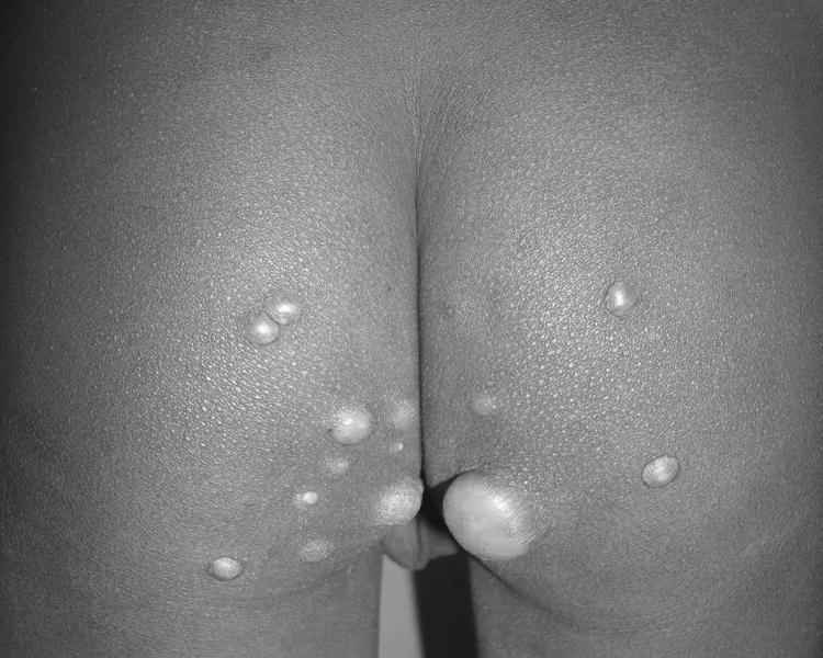
Clinical photograph of the child showing molluscum contagiosum of various sizes over the buttocks.
2. Discussion
The appearance of MC lesions in adult men requires an evaluation of the immunocompromised state, but in children MC is rarely associated with immunodeficiency and usually no further evaluation is needed [3]. In this case report, the importance of clinical suspicion, even in the absence of a definite history, is highlighted. The child was apparently asymptomatic without any systemic illness. The death of the parents was a clue to the diagnosis. Probably, the infection was acquired through the vertical route from the mother. The awareness of this condition as being the first sign of HIV infection should prompt the diagnostic investigation, especially in the countries with limited access to healthcare facilities.
In HIV-positive patients, MC tends to occur during the advanced phase of the disease and signifies advancing immunosuppression [3]. Moreover, in HIV patients, lesions can be large, and mimic cutaneous tumors [4]. Genital lesions are especially common in sexually active individuals. In this case, no history of sexual abuse could be elicited. Living in close proximity, skin-to-skin contact, sharing of fomites, and residing in tropical climates increases the spread of infection [5]. Diagnosis is made on the distinctive clinical appearance, but can be confirmed by skin biopsy and uncommonly by stained smears of expressed cores. Warts, varicella, intradermal nevi, lichen planus, and basal cell carcinomas are included in the differential diagnosis [2].
Even in immunocompetent individual’s treatment of MC is recommended because of the high rate of associated symptoms, risk for transmissibility, and cosmetic or social concerns. Major therapeutic modalities employed for MC are physical destruction, immunomodulation, and antiviral agents. Combinations of these therapies may be employed [6]. Lesions can be destroyed by the application of cantharidin, cryotherapy and curettage, topical tretinoin and topical imiquimod 5% cream. Topical imiquimod 5% cream applied 3 times a week is the preferred therapy for MC lesions localized to the anogenital area [7]. MC is difficult to treat in patients with HIV. Optimizing antiretroviral therapy and using lesion-destroying therapies are usually helpful.
References
Cite this article
TY  - JOUR
AU  - Sriparna Basu
AU  - Ashok Kumar
PY  - 2013
DA  - 2013/07/16
TI  - Giant Molluscum contagiosum – A clue to the diagnosis of Human Immunodeficiency Virus infection
JO  - Journal of Epidemiology and Global Health
SP  - 289
EP  - 291
VL  - 3
IS  - 4
SN  - 2210-6014
UR  - https://doi.org/10.1016/j.jegh.2013.06.002
DO  - 10.1016/j.jegh.2013.06.002
ID  - Basu2013
ER  -