
2.4 Aortofemoral Plethysmographic Volume Wave Velocity Obtained During the Routine 12 Channel ECG Corresponds in its Determinants to Tonometrically Derived Carotid-Femoral Pulse Wave Velocity
- DOI
- 10.2991/artres.k.191224.009How to use a DOI?
- Abstract
Background: The lack of acceptance in clinical routine represents a major obstacle for carotid-femoral pulse wave velocity (cfPWV) measurements. We thought to include PWV measurements into the 12-channel ECG, which is performed routinely.
Methods: Using the conventional electrode position for the 12 channel ECG, arterial impedance plethysmographic signals were obtained from the four extremities at 40 kHz [1]. It was confirmed that the obtained pulse synchronous volume waves originate at the level of the elbow and the knee. In analogy to cfPWV measurements, the volume wave velocity at the aorta and femoral arteries (VWVtorso) was calculated from the time differences of the plethysmographic signals between arms and legs.
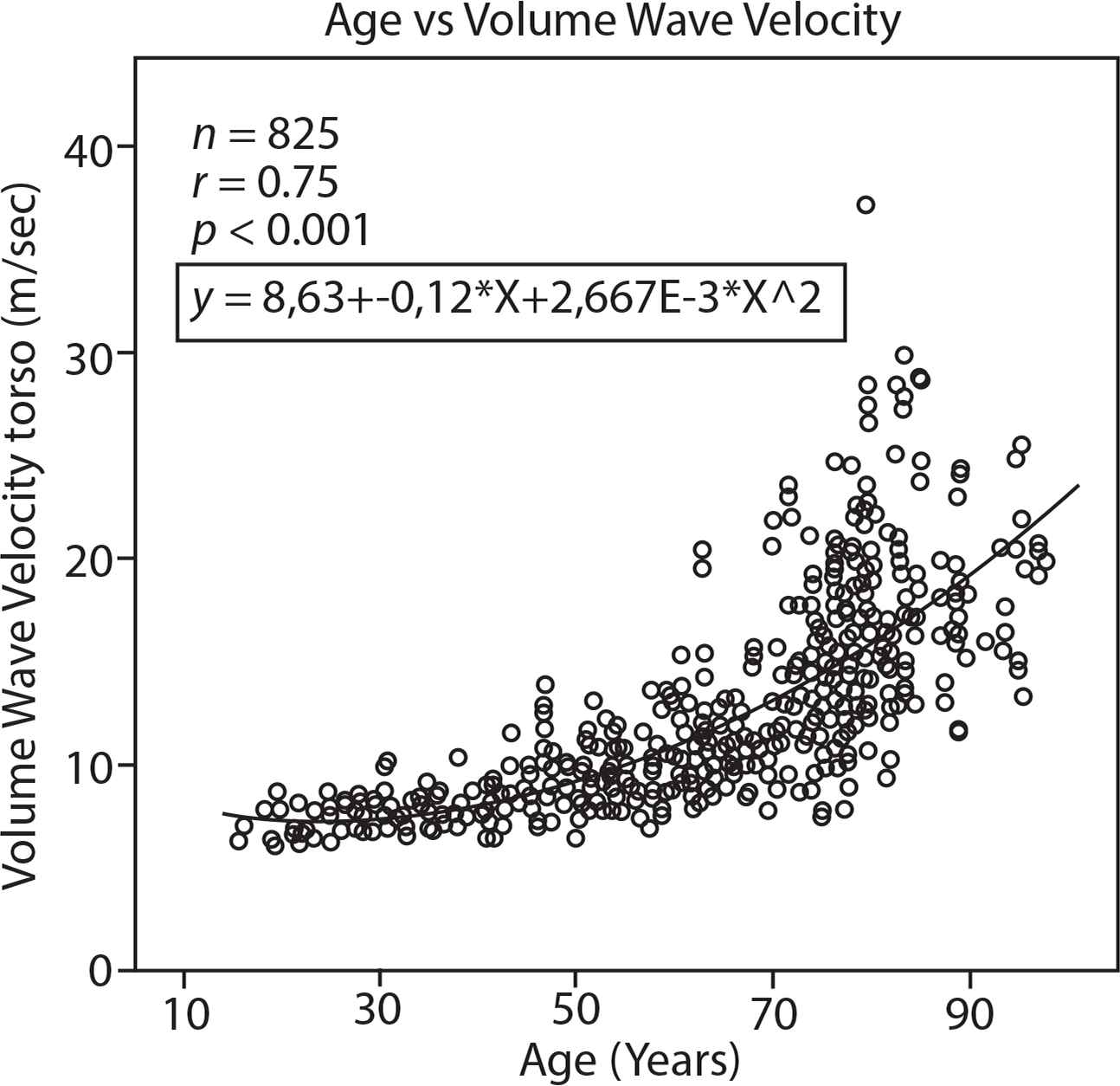
Results: Transit times of R-wave to volume wave at the leg were longer but closely related to tonometrically derived transit times (199 ± 26 vs 134 ± 24 m/sec, n = 115, r = 0.93, p < 0.001). VWVtorso in 115 participants was higher than tonometrically determined cfPWV, (13.2 ± 5.81 vs 8.8 ± 2.98, ± SD, p < 0.001), since muscular arteries are included in VWVtorso. VWVtorso was measured in 825 healthy participants and patients with cardiovascular diseases (aged 16 to 97 years) (Figure 1). In stepwise multiple regression analysis, VWVtorso was found to be related positively to known cardiovascular risk factors such as age, blood pressure, HbA1C and LDL-cholesterol (p < 0.001 for all) and also negatively to appendicular muscle mass [2]. VWVtorso corresponds in its physiological determinants closely to those of cfPWV.
Conclusion: These background arterial impedance plethysmographic measurements yielding VWVtorso, made without time delay during routine 12 channel ECG, show promise for large scale, routine clinical assessment of large artery function.
- Copyright
- © 2019 Association for Research into Arterial Structure and Physiology. Publishing services by Atlantis Press International B.V.
- Open Access
- This is an open access article distributed under the CC BY-NC 4.0 license (http://creativecommons.org/licenses/by-nc/4.0/).
Download article (PDF)
View full text (HTML)
Cite this article
TY  - JOUR
AU  - Falko Skrabal
AU  - Thomas Weber
AU  - Katharina Skrabal
PY  - 2020
DA  - 2020/02/15
TI  - 2.4 Aortofemoral Plethysmographic Volume Wave Velocity Obtained During the Routine 12 Channel ECG Corresponds in its Determinants to Tonometrically Derived Carotid-Femoral Pulse Wave Velocity
JO  - Artery Research
SP  - S11
EP  - S12
VL  - 25
IS  - Supplement 1
SN  - 1876-4401
UR  - https://doi.org/10.2991/artres.k.191224.009
DO  - 10.2991/artres.k.191224.009
ID  - Skrabal2020
ER  -