
P.20 Evolving Structure-Function Correlates during Aortic Maturation and Aging
- DOI
- 10.2991/artres.k.201209.033How to use a DOI?
- Keywords
- Microstructure function; mechanics cells
- Abstract
Introduction: Unraveling aortic cellular and extracellular microstructural and mechanical mechanisms triggered to maintain homeostasis in murine aortae during maturation and aging is fundamental to better understand remodeling in human arteriopathies [1].
Methods: This study, combining ex-vivo extension-inflation testing [2], multiphoton microscopy and optical histology, aimed to quantify multiple microstructural parameters of primary extracellular components – collagen, elastic lamellae – and cells – endothelial, smooth muscle and adventitia cells – of the aorta with a dynamic and multiregional 3D approach. The analysis focused on the quantification and correlation of the histo-mechanical properties of the thoracic aorta as a function of age from 21 days to 1 year after birth, that is, from the time of weaning to maturation and therefore the natural aging. The parameters quantifying the three-dimensional microstructural phenomena of deposition, remodeling and removal of aortic components under pressure and stretch conditions equivalent to those in vivo were layer thicknesses, straightening, alignment and thickness of collagen bundles, number and size of elastic lamellae, density and alignment of the different vascular cells.
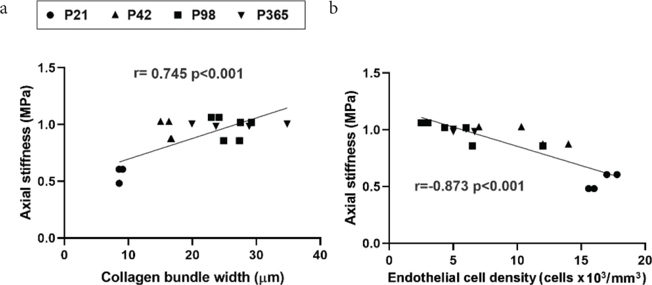
Results: Changing dynamics at different ages were characterized, such as the smooth muscle cell population reduction and hypertrophy with the interlamellar widening from an intermediate age. Significant correlations indicated the fundamental role of both cells and deposited extracellular proteins such as the reduction in endothelial and smooth muscle cell densities but also the increase in straightness and thickness of collagen bundles in relation to the increase in circumferential and axial stiffness of the aortic wall.
 Figure
Microstructure-mechanics correlates.
- Copyright
- © 2020 Association for Research into Arterial Structure and Physiology. Publishing services by Atlantis Press International B.V.
- Open Access
- This is an open access article distributed under the CC BY-NC 4.0 license (http://creativecommons.org/licenses/by-nc/4.0/).
Cite this article
TY  - JOUR
AU  - Cristina Cavinato
AU  - Jay D Humphrey
PY  - 2020
DA  - 2020/12/31
TI  - P.20 Evolving Structure-Function Correlates during Aortic Maturation and Aging
JO  - Artery Research
SP  - S42
EP  - S42
VL  - 26
IS  - Supplement 1
SN  - 1876-4401
UR  - https://doi.org/10.2991/artres.k.201209.033
DO  - 10.2991/artres.k.201209.033
ID  - Cavinato2020
ER  -