
Supervised Filter Learning for Coronary Artery Vesselness Enhancement Diffusion in Coronary CT Angiography Images
- DOI
- 10.2991/ijcis.d.200422.001How to use a DOI?
- Keywords
- Computed tomography angiography; Coronary artery filtering; Vesselness enhancement; Machine learning; Vessel segmentation
- Abstract
In medical imaging, vesselness diffusion is usually performed to enhance the vessel structures of interest and reduce background noises, before vessel segmentation and analysis. Numerous learning-based techniques have recently become very popular for coronary artery filtering due to their impressive results. In this work, a supervised machine learning method for coronary artery vesselness diffusion with high accuracy and minimal user interaction is designed. The fully discriminative filter learning method jointly learning a classifier the weak learners rely on and the features of the classifier is developed. Experimental results demonstrate that this scheme achieves good isotropic filtering performances on both synthetic and real patient Coronary Computed Tomography Angiography (CCTA) datasets. Furthermore, region growing-based segmentation approach is performed over filtered images obtained by using different schemes. The proposed diffusion scheme is able to achieve higher average performance measures (87.8%
- Copyright
- © 2020 The Authors. Published by Atlantis Press SARL.
- Open Access
- This is an open access article distributed under the CC BY-NC 4.0 license (http://creativecommons.org/licenses/by-nc/4.0/).
1. INTRODUCTION
Accurate coronary arteries determination is commonly a fundamental step for computer-aided diagnosis of cardiovascular diseases, especially for coronary stenosis quantification [1]. In clinical practice, medical imaging techniques-based vessel detection usually contains two significant tasks, namely vascular structures enhancement in original medical images and vessel segmentation. In the literature, a variety of automatic two-dimensional (2D) or three-dimensional (3D) vessel segmentation algorithms have been proposed. For example, 3D coronary arteries can be directly segmented based on grayvalue [1], prior knowledge [2,3], deformable model [4] and learning-based method [5].
In Kerkeni et al. [6], the author compares four Hessian-based multiscale filters and shows that the vessel enhancement diffusion (VED) filter is superior to the other two methods in enhancing vascular structure and suppressing background noise. At present, there are three main advantages of VED. First, VED can improve the separation of blood vessels in maximum intensity projection (MIP) because the intensity distribution of each filter vessel becomes more uniform. In addition, strong diffusion in the vessel direction helps to overcome significant intensity decline. Thirdly, VED can inhibit nonvessel structures. Despite such a large number of vessel enhancement and segmentation methods, robust enhancement of 3D curved structures is still a challenging task, due to the intensity variations along the 3D coronary artery structures and the existence of surrounding noises and tissues.
In many cases, handcrafted image filters often require complex mappings, and the underlying models of the imaging processes are not always easily obtained or constructed. Therefore, neural networks can be used for function approximation and the data modeling process [7]. Among these methods, the The Deep Convolution Network [8] has recently become very popular due to its impressive results on many reference datasets [9]. Generally, large convolutional networks are trained by back propagation to minimize classification errors. However, due to the large number of parameters to be optimized, deep network needs a lot of training data and computing power to achieve the latest performance. When the input image is too large or only a limited number of training data are available (this is a typical case in the biomedical field), their applicability and performance will be reduced. Furthermore, the neural network architecture used in [8] needs special settings and careful design, and even on GPU, training is expensive in computing. In contrast, our method requires much less computation and involves only a few parameters that are easy to adjust. Moreover, it enables us to process 3D medical image stacks, which, despite their prominent position in the medical field, due to its low computational burden. The basic idea of designing a neural network image filter is to construct a function in a neural network to map an image on another image in which certain properties of the image are extracted or enhanced.
Our goal is to design a supervised machine learning method for coronary artery vesselness diffusion, with high accuracy and minimal user interaction. Like our method, several authors have used cascading of classifiers [10–14] to improve the performance of classification problem. These classifiers use a set of features extracted from the output of the previous layer for training. Since each classifier is treated separately, the training process is relatively easy. We use an iterative process for regression. Generally, in such a framework, nonlinear transformation is sequentially applied to the input signal to obtain a higher-order representation of the input, capture more context information and improve the classification rate in each iteration. We compute the kernel in a closed form, which allows us to handle large parameter spaces. Our method is easier to train by supervised optimization of features and continuous regression variables one by one. In addition, it can effectively handle a large number of inputs.
This paper is the extension of our previous conference work [15]. The scope of this work is in the first place to investigate and analyze the abilities of deep neural networks for medical image filtering. The main contributions are summarized in the following aspects: (1) A detailed review of related work about learning-based coronary artery vesselness diffusion in CCTA images is presented. (2) A fully discriminative filter learning method jointly learning a classifier our weak learners rely on and the features of the classifier is developed. Compared to handcrafted image filters, neural network image filters are designed and trained for edge extraction, vesselness enhancement and noise suppression. (3) The theoretical supports of the Gradient Boosting framework and its quadratic approximation used in this work are presented.
In the following sections, the remaining part of this work is presented: Section 2 presents the proposed coronary artery vesselness filter learning approach, which includes three parts: (1) general introduction about gradient boosting algorithm; (2) formation of regression trees and decision stumps to optimize the Gradient Boosting classifier; (3) convolution kernels learning while building the regression trees. Section 3 presents the experimental results on public datasets, and real patient CCTA datasets from the hospital. Section 4 gives a brief discussion. Finally, conclusions are given in Section 5.
2. MATERIALS AND METHODS
2.1. Gradient Boosting
The boosting motivation is the process of combining the output of many weak classifiers to produce a powerful combination, which is a way to fit the addition expansion into a set of basic functions. In general, these models are fitted by minimizing the loss functions averaged over the training data [16,17,18].
Gradient Boosting is usually used to approximate the function
Now consider the training samples
For the above minimization problem, it does not exist a general solver. However, a regression weak learner or learning algorithm, denoted as
For twice differentiable loss functional
Therefore, optimizing the loss function
Algorithm 1: Gradient Boosting framework with quadratic approximation
Input:
let
for k = 0, 1, 2, … do
let
either
or
let
pick
let
choose step size
let
end for
2.2. Regression Trees
Gradient Boosting algorithm is typically implemented by searching through the collections of weak learners that are dependent on a fixed set of features. Starting from the root, the regression tree learning procedure is learned by building one split at a time, as described in [17]. Then
By differentiating
Typically, a split is comprised of a test function
At iteration
Then a test function
The whole minimization procedure is performed in stages. The proposed approach is described in the following aspects: First a set of kernel candidates are constructed. For each candidate, the optimal value of
2.3. Convolution Kernels
In this work, we let kernels k to be square windows within x, in the order to make the minimization computations tractable. Now consider a square window centered at
To avoid overfitting problem, we first introduce the regularization term, in order to produce smooth kernels. Then Eq. (11) becomes
Secondly, filters and splits are learned on a subset of random samples from the training set. When learning the rank functions, we pick
Then we pick
Then the split cost is
By computing the lowest split cost
Three three-dimensional (3D) examples of synthetic vessel models from the March 2013 VascuSynth Sample: Group 2 (data3), Group 3 (data4) and Group 4 (data5).
2.4. Segmentation
A variety of research efforts in the literature for 3D coronary arteries segmentation from CCTA datasets have been studied. Existing 3D coronary arteries segmentation methods can be divided into two main classes: deformable models [20–22] and differential measures [23]. For the purpose of evaluating the performances of different diffusion filters, each real patient CCTA dataset was first filtered by different diffusion algorithms. Complete coronary artery structures were then segmented from different filtered CCTA images based on the region growing method [24,25]. The region growing-based method in this work includes three main steps. First, a global threshold is set to around gray intensity of 200-300 if the lowest CT intensity is −2048, or set as 400-500 if the lowest CT intensity is −1024, which will give a roughly segmented 3D region. To obtain accurate local region, a 3D mask is necessary. Region growing can use mask as a parameter. The main purpose of local region is let the threshold or region growing to be restricted to one branch or several branches of vessel. Second, irrelevant regions need to be removed. After the removal of other tissue, the skeleton similar structure was extracted. Finally, from the triangle faces, the vertices or faces normal are computed and used as an orientation for further region growing. The theory of the growing is based on image profile or maximal gradient along normal. Each complete coronary artery segmentation result was compared to the ground-truth region. The ground truth coronary arteries regions for all four CCTA datasets were labeled by the experienced radiologist from the National Heart Center Singapore. All the parameters and threshold values of region growing segmentation method were kept fixed for each diffusion filter for a fair comparison.
3. RESULTS
The experimental results presented includes two parts. Firstly, the proposed method is tested on VascuSynth Sample database and compared with Cheng's method [26]. Secondly, different diffusion schemes are tested on the CCTA images from real patients, to evaluate the segmentation accuracy of the proposed diffusion scheme.
3.1. Validation on Publicly Available Database
In this stage, the segmentation approach is tested on the March 2013 VascuSynth Sample (10 datasets) presented in [26]. As reported in [26], four different quantitative measures for the synthetic validation, i.e., true positives (TP), false negatives (FN), false positives (FP) and overlap measure (OM) between the obtained vessel segmentation and the ground truth vessel segmentation. Figure 1 presents three examples of synthetic vessel models from the March 2013 VascuSynth Sample: Group 2 (data3), Group 3 (data4) and Group 4 (data5), which are used as the ground truth vessel structures and adequate to represent the 3D vascular structures, for the purpose of evaluating the efficiency of the proposed vesselness filter function. Table 1 summarizes average TP, FN, FP and OM rates on the 10 datasets. Moreover, 3 datasets on the 2011 VascuSynth Sample Data are used in the noise experiments to compare with the Cheng's method. Low level Gaussian noise was added to the 2011 VascuSynth Sample Data (3 datasets) [26], and the proposed method was tested compared with Cheng's method [26]. The comparison results are presented in Table 2. It can be seen that the proposed method slightly outperforms Cheng's method with the presence of low level Gaussian noise.
| Data Set | TP | FN | FP | OM |
|---|---|---|---|---|
| 1 | 97.94 | 2.06 | 3.63 | 97.90 |
| 2 | 96.02 | 3.98 | 6.48 | 95.24 |
| 3 | 95.43 | 4.57 | 7.02 | 94.58 |
| 4 | 94.87 | 5.13 | 7.71 | 94.29 |
| 5 | 97.60 | 2.40 | 4.09 | 96.81 |
| 6 | 96.88 | 3.12 | 5.98 | 96.13 |
| 7 | 97.26 | 2.74 | 5.05 | 96.82 |
| 8 | 96.19 | 3.81 | 6.39 | 95.40 |
| 9 | 97.25 | 2.75 | 4.67 | 97.06 |
| 10 | 97.33 | 2.67 | 5.54 | 97.04 |
| Avg. |
96.67 |
3.32 |
5.66 |
96.13 |
TP, true positives; FN, false negatives; FP, false positives; OM, overlap measure.
Segmentation results on 2013 VascuSynth sample (%).
| TP |
FN |
FP |
OM |
|||||
|---|---|---|---|---|---|---|---|---|
| Cheng's | Ours | Cheng's | Ours | Cheng's | Ours | Cheng's | Ours | |
| Data1 | 94.38 | 95.54 | 5.62 | 4.46 | 7.49 | 6.93 | 93.83 | 94.16 |
| Data2 | 94.87 | 95.06 | 5.13 | 4.94 | 5.38 | 4.62 | 94.55 | 94.79 |
| Data3 | 95.29 | 96.07 | 4.71 | 3.93 | 5.63 | 4.95 | 94.89 | 95.71 |
TP, true positives; FN, false negatives; FP, false positives; OM, overlap measure.
Comparison of Cheng's method with our method in the presence of low level of gaussian noise (σ2 = 20) (%).
3.2. CT Images Diffusion
The proposed diffusion scheme was further applied into four CCTA datasets in DICOM format, implemented on a 16 GB of RAM Windows laptop. The parameters in this part are chosen as total diffusion time 15s and time step 0.25s. The proposed diffusion scheme is proven to be efficient, with an average processing time of about 14.5 minutes. This enables the proposed diffusion scheme to be further applied as a real-time CCTA images preprocessing tool in clinical practice.
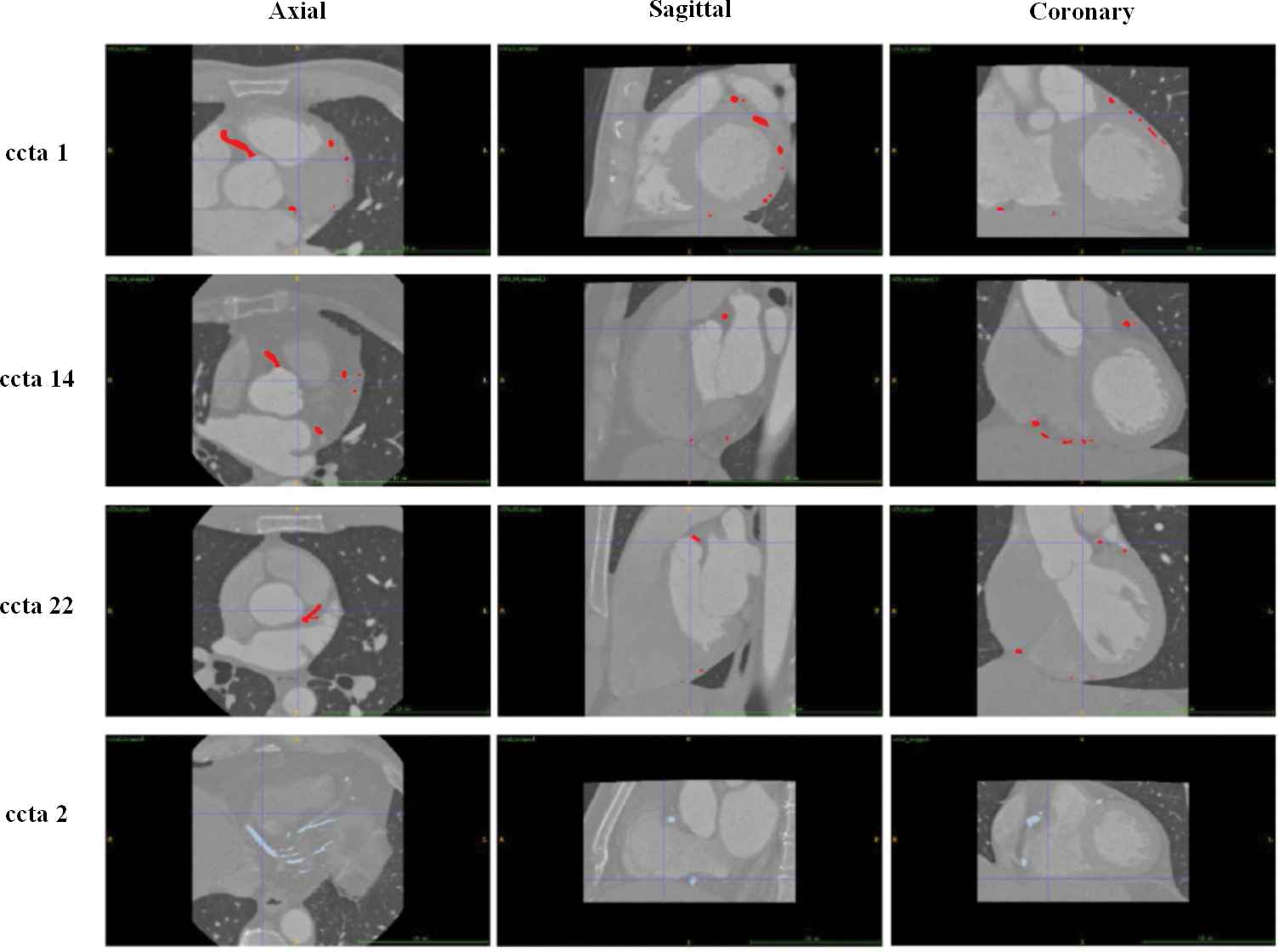
Segmentation results in axial, sagittal and coronary view for ccta1, ccta14, ccta 22 and ccta 2.
Once the original CCTA datasets were filtered by the proposed diffusion scheme, region growing-based segmentation method was applied to segment the coronary arteries. Figure 2 depicts the segmentation results obtained by performing the proposed vesselness diffusion scheme in axial, sagittal and coronary view for all four CCTA datasets. The segmented results were compared to the radiologist labeled ground-truth regions in three different views, and were proven to highly overlap with the ground-truth regions, which demonstrated the robustness of the proposed diffusion scheme. On the other hand, segmentation results without vesselness diffusion were observed to poorly overlap with the ground-truth regions. Many surrounding noises and isolated points were segmented, and many thin vessel branches were even missing. Moreover, a comparison of our segmentation results (red) with the ground truth regions (blue) in axial view for ccta1, ccta14, ccta22 is presented in Figure 3.
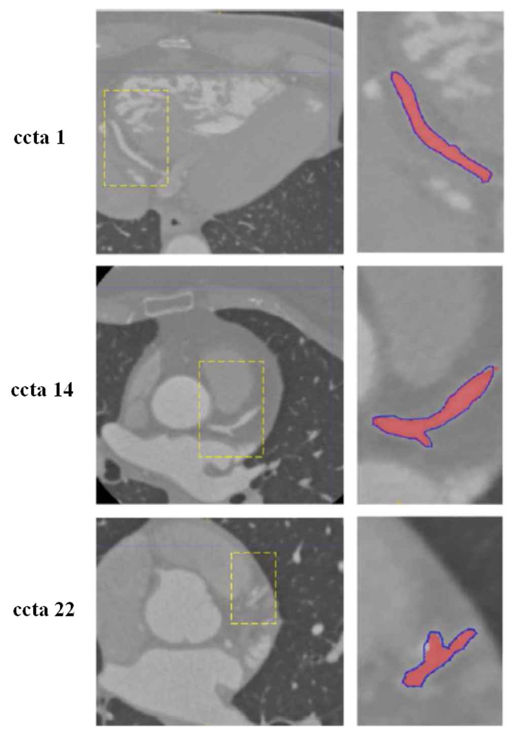
A comparison of our segmentation results (red) with the ground truth regions (blue) in axial view for ccta1, ccta14 and ccta 22.
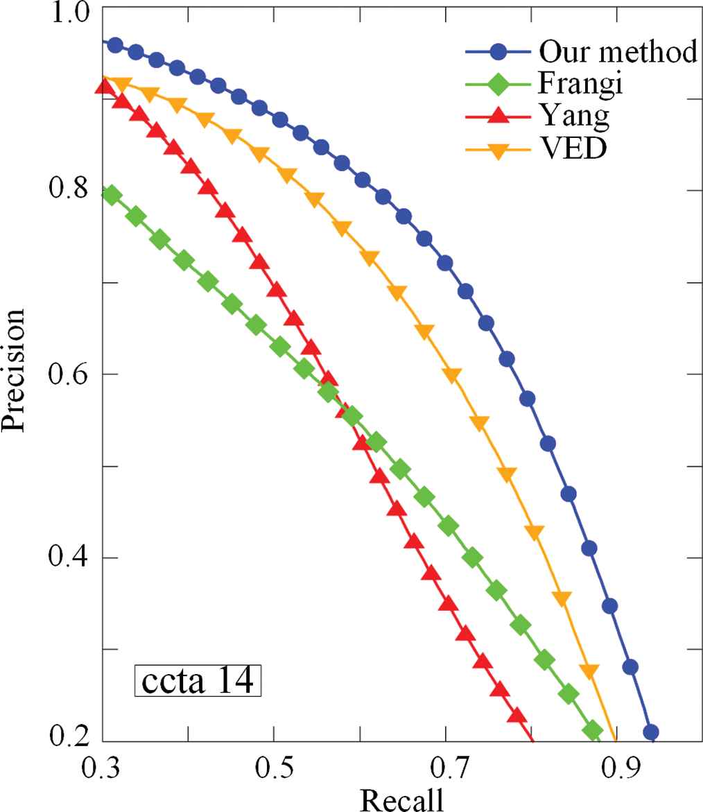
Precision-recall curves for voxel classification over ccta 14, obtained for different thresholds on
| ccta 1 |
ccta 2 |
ccta 14 |
ccta 22 |
|||||||||
|---|---|---|---|---|---|---|---|---|---|---|---|---|
| Dice | Precision | Sensitivity | Dice | Precision | Sensitivity | Dice | Precision | Sensitivity | Dice | Precision | Sensitivity | |
| Frangi | 0.82 | 0.83 | 0.80 | 0.77 | 0.84 | 0.76 | 0.79 | 0.80 | 0.78 | 0.80 | 0.79 | 0.81 |
| Yang's | 0.84 | 0.84 | 0.83 | 0.80 | 0.84 | 0.78 | 0.79 | 0.82 | 0.77 | 0.78 | 0.81 | 0.83 |
| VED | 0.85 | 0.85 | 0.87 | 0.82 | 0.86 | 0.85 | 0.81 | 0.83 | 0.79 | 0.82 | 0.83 | 0.85 |
| Proposed | 0.89 | 0.93 | 0.92 | 0.91 | 0.93 | 0.92 | 0.87 | 0.86 | 0.87 | 0.88 | 0.89 | 0.91 |
CCTA, Coronary Computed Tomography Angiography; VED, vessel enhancement diffusion.
A comparison of three performance measures (Dice, Precision and Sensitivity) of different diffusion methods for all four CCTA datasets.
Furthermore, to explore the performance of the proposed diffusion scheme, three other diffusion methods (Frangi's, Yang's [27] and VED) were also performed into four CCTA datasets. The same region growing method was then applied to segment the complete coronary arteries, to compare with the ground-truth results. To compute the probability of each pixel in the input image of being part of a vessel structure
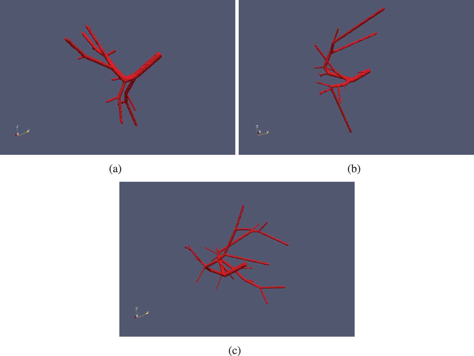
Finally, in Figure 5 we show the coronary arteries rendering results by the proposed diffusion scheme. It can be observed that main branches of coronary arteries can be completely detected. The vessels are found to be enhanced and connected, and no nonvessel tissues and artifacts are connected to the coronary artery vascular structures. Additionally, the new method is capable of extracting even small vessel branches. Experimental results demonstrate that the proposed diffusion filter can effectively reduce the pseudo coronary artery structures and isolated noisy points.
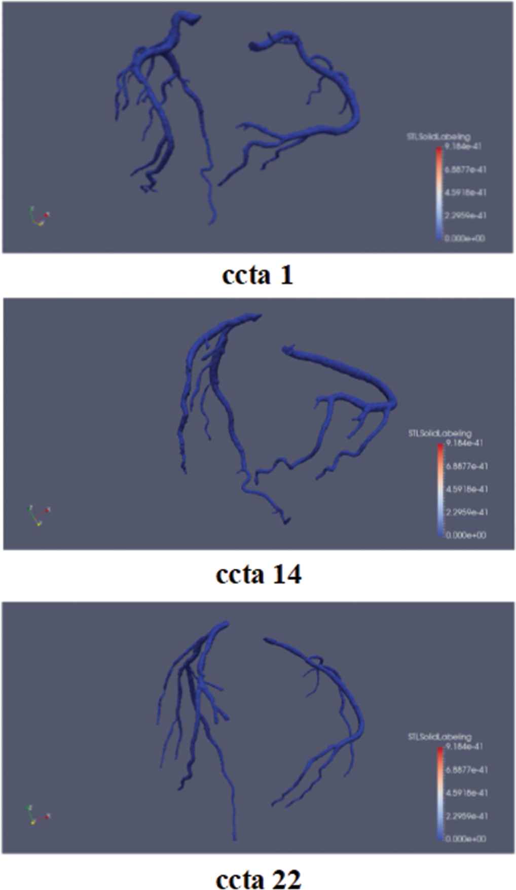
Coronary arteries rendering results by the proposed diffusion scheme for ccta 1, ccta 14 and ccta 22.
4. DISCUSSION
In this work, a detailed review of related work about learning-based coronary artery vesselness diffusion in CCTA images is presented. Besides, a fully discriminative filter learning method jointly learning a classifier our weak learners rely on and the features of the classifier is developed. Thirdly, the theoretical supports of the Gradient Boosting framework and its quadratic approximation used in this work are presented. The developed filter learning method is found to outperform the current latest learning-based segmentation techniques. Moreover, it ensures that almost no parameter adjustment is needed during training and testing procedure, which avoids parameter and feature selection tasks. Apart from using the public datasets, the proposed algorithm is evaluated on a total of four sets of CCTA images from the hospital, which ensures enough statistical sampling.
In the future, more real patient CCTA datasets with ground truth labels will be obtained from the hospitals and related research agencies. Besides, different kinds of data argumentation operations will also be performed before training the algorithm. We believe that the performance of the algorithm can be further improved with enough training datasets. Furthermore, different kinds of neural networks, which utilize multi-view or multi-modality information will be further developed, for the purpose of improving the network's ability of segmenting coronary arteries from CCTA images. On the other hand, validation results on real patient CCTA datasets are based on the manually measured sequence of lumen area. Due to the existence of coronary artery stenosis, calcium, plaques and surrounding noises, the measured region of interest varies for different experts. Later for ground truth lumen contour labeling, we may need at least two professional cardiologists.
5. CONCLUSION
In this work, we propose an accurate and efficient learning-based vesselness filtering scheme, for the purpose of enhancing coronary arteries and reducing background noises in CCTA images. The fully discriminative filter learning method jointly learning a classifier our weak learners rely on and the features of the classifier. Moreover, the theoretical supports of the Gradient Boosting framework and its quadratic approximation used in this work are presented. Experimental results demonstrate that the new 3D anisotropic diffusion scheme outperform the standard and Kroon's optimized scheme for the task of curve-like features enhancement and noise reduction in the synthetic datasets. Quantitative results on VascuSynth Sample indicate that the proposed method is slightly better than Cheng's method for small or thin vessels. Moreover, compared to the existing diffusion methods like Frangi's, improved Hessian and VED, our proposed method shows strong enhancement for coronary arteries and resists to noisy background tissues in real patient CCTA images. More specifically, it is observed that more thin vessel branches can be detected and less noisy structures are introduced in the coronary artery results segmented from the CCTA images filtered by the proposed diffusion scheme. Compared with the human labeled ground-truth coronary arteries, the segmentation results by performing the proposed diffusion scheme can achieve higher OMs (87.8%
CONFLICT OF INTEREST
The authors declare that they have no conflict of interest.
AUTHORS' CONTRIBUTIONS
H.F. Cui designed all the experiments and prepared the manuscript.
ACKNOWLEDGMENTS
The study was supported in part by the National Natural Science Foundation of China under Grant 61801393, and in part by the Natural Science Basic Research Project in Shaanxi of China (Program No. 2019JQ-254). The authors thank the National Heart Centre Singapore for the real-patient CCTA datasets.
REFERENCES
Cite this article
TY  - JOUR
AU  - Hengfei Cui
PY  - 2020
DA  - 2020/05/06
TI  - Supervised Filter Learning for Coronary Artery Vesselness Enhancement Diffusion in Coronary CT Angiography Images
JO  - International Journal of Computational Intelligence Systems
SP  - 488
EP  - 495
VL  - 13
IS  - 1
SN  - 1875-6883
UR  - https://doi.org/10.2991/ijcis.d.200422.001
DO  - 10.2991/ijcis.d.200422.001
ID  - Cui2020
ER  -