
Occlusal characteristics and prevalence of associated dental anomalies in the primary dentition
- DOI
- 10.1016/j.jegh.2014.07.001How to use a DOI?
- Keywords
- Primary teeth; Occlusion; Gemination; Fusion; Hypodontia; Cross-bite
- Abstract
Introduction: Morphological variations in primary dentition are of great concern to a pediatric dentist as it may pose clinical problems like dental caries, delayed exfoliation and also anomalies in the permanent dentition, such as impaction of successors, supernumerary teeth, permanent double teeth or aplasia of teeth. The present study was conducted to investigate the presence of dental anomalies in the primary dentition of 1000 schoolchildren in the 3–5 year-old age group in Faridabad.
Materials and methods: One-thousand schoolchildren were examined using Type III examination (WHO, 1997) for primary molar relationship, occlusal characteristics, primate spaces, physiological spaces and other anomalies of teeth, including number and morphology.
Results and conclusions: The prevalence of physiological spaces in maxillary and mandibular arches was 50.9% and 46.7%, respectively, whereas primate spaces were found in 61.7% of the children in the maxillary arch and 27.9% in the mandibular arch. The prevalence of unilateral anterior and posterior cross-bite was 0.1% and 0.8%, respectively, in the present study. The prevalence of hypodontia in the primary dentition was found to be 0.4% and the prevalence of fusion and gemination in the present study was 0.5%. Double teeth (fusion and gemination) and hypodontia were the most common dental anomalies found in the primary dentition in the present study.
- Copyright
- © 2014 Ministry of Health, Saudi Arabia. Published by Elsevier Ltd.
- Open Access
- This is an open access article under the CC BY-NC-ND license (http://creativecommons.org/licenses/by-nc-nd/4.0/).
1. Introduction
Occlusion in primary dentition plays a significant role in guiding the occlusion in succeeding permanent dentition [1,2]. The characteristic set of features in primary dentition to a large extent lays the foundation for proper eruption and alignment of the succeeding dentition. Based on the observation of these key features of occlusion in the child’s dentoalveolar system during the formative years, the characteristics of the permanent dentition/occlusion can be predicted very well [3].
Morphological variations in primary dentition are of great concern to a pediatric dentist as they may pose clinical problems associated with them, including dental caries, delayed exfoliation, and also anomalies in the permanent dentition, such as impaction of successors, supernumerary teeth, permanent double teeth, or aplasia of teeth [4]. Thus, early diagnosis of these anomalies allows for more comprehensive long-term treatment planning, favorable prognosis and less extensive interception [5,6].
The occlusal traits in the primary dentition vary among different populations and ethnic groups [7]. Therefore, the present study was conducted on 1000 3–5 year-old schoolchildren attending school in Faridabad city to investigate the presence of dental anomalies in the primary dentition. The objectives of this study were to assess the characteristic features of occlusion in primary dentition with the following parameters:
- ▪
To assess the terminal molar relations on the right and left side of the jaw;
- ▪
To assess the presence of any anterior or posterior cross-bite;
- ▪
To assess the presence of scissor-bite;
- ▪
To assess the presence of infra-occlusion;
- ▪
To assess the presence of any other tooth anomalies, including number and morphology.
2. Materials and methods
The present study was conducted on 3–5 year-old school children in Faridabad city, Haryana. Ethical clearance was sought from the ethics committee of Sudha Rustagi College of Dental Sciences and Research, Faridabad, after explaining the aim and importance of the study. The training and calibration of the examiner was done under the guidance of a supervisor in the Department of Pedodontics, Sudha Rustagi College of Dental Sciences and Research, Faridabad, to limit the intra-examiner variability. The training and calibration was repeated until the examiner produced consistent results. The study was conducted on school premises after obtaining written permission from school authorities and written informed consent from parents that were distributed to children two days prior to the visit of the examiner.
The following criteria were used to select the children who participated in this study:
Inclusion criteria of the study:
- 1.
Children with a complete set of primary dentition without premature loss of primary teeth.
- 2.
Children with no erupted permanent teeth.
- 3.
Children with teeth free from caries.
- 4.
Children whose parents gave written informed consent.
Exclusion criteria of the study:
- 1.
Grossly decayed anterior teeth and molars.
- 2.
Children with any systemic disease.
- 3.
Children with un-cooperative behavior.
The study was carried out in a classroom provided by the school authorities. The clinical examination was conducted in natural light using a mirror and a probe. The subjects were examined using Type III clinical examination (WHO, 1997), and observations were recorded. A total of 1000 children aged 3–5 years from selected schools finally formed the sample population.
The following parameters were recorded:
- 1.
Primary molar relationship: The mesiodistal relationship between the distal surfaces of the upper and lower second deciduous molars were recorded according to the Baume (1950) classification:
- (a)
Flush terminal plane
- (b)
Distal step
- (c)
Mesial step
- (d)
Asymmetrical molar relation
- (a)
- 2.
Anterior cross-bite
- 3.
Posterior cross-bite
- 4.
Infra-occlusion
- 5.
Scissors bite
- 6.
Tooth anomalies: Presence of tooth anomaly was also recorded, i.e., Anodontia, Hypodontia, Oligodontia, Hyperdontia, Supplemental, Supernumerary, Transposition, Microdontia, Macrodontia, Fusion, Talon’s cusp, Ectopic eruption, Dens evaginatus or any other.
The obtained data were coded and entered in Microsoft Excel which was then subjected to statistical analysis. Categorical data were analyzed by Chi-square test for differences between groups. Significance for all statistical tests was predetermined at a probability value of 0.05 or less (data were analyzed using the statistical package SPSS Software, Version 14, Chicago, USA).
3. Results and discussion
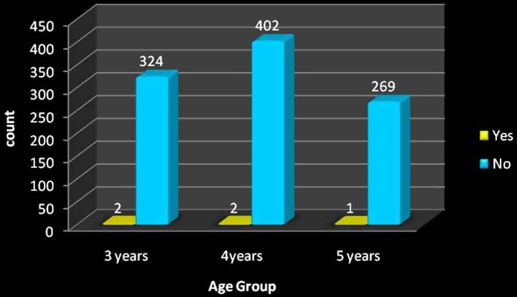
The present study was conducted on 1000 schoolchildren aged 3–5 years of Faridabad city, Haryana. Table 1 shows the distribution of examined children according to age and sex. Out of 1000 children, 326 (32.6%), 404 (40.4%), and 270 (27%) were 3, 4 and 5 years of age, respectively. The study consisted of 53.3% males and 46.7% females. According to the Pearson Chi-Square test, the age-wise distribution of both the sexes (male and female) was not found to be statistically significant (p-value: 0.070).
| Age | Sex | Total | |
|---|---|---|---|
| M | F | ||
| 3 years | 165 | 161 | 326 |
| 50.60% | 49.40% | ||
| 4 years | 208 | 196 | 404 |
| 51.50% | 48.50% | ||
| 5 years | 160 | 110 | 270 |
| 59.30% | 40.70% | ||
| Total | 533 | 467 | 1000 |
| 53.30% | 46.70% | ||
| Pearson Chi-square | 5.332 | ||
| p-value | 0.07 | ||
Percentage distribution of children according to age and sex.
The flush terminal plane relationship of deciduous second molars was noted in 65.1% of the children followed by 12.8% (mesial step relationship) and 2.4% (distal step relationship). The asymmetric molar relationship (left and right molar relations not the same) was seen in 18.8% of the children. The distribution of physiological and primary spaces, anterior and posterior cross-bites, hypodontia, fusion, and gemination is also depicted in Figs. 1–5.
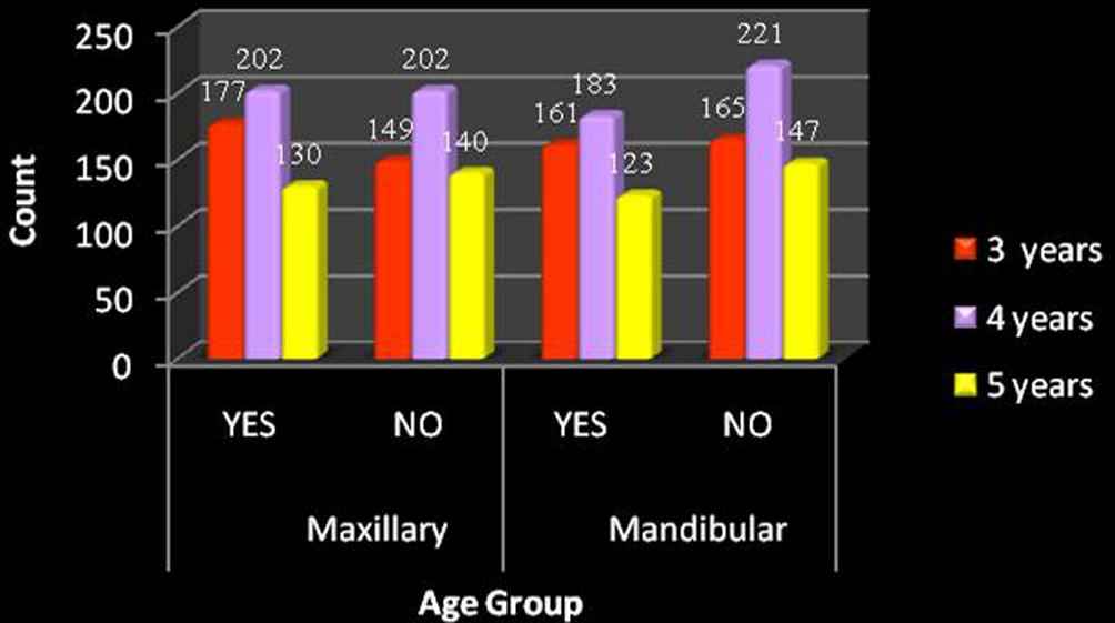
Age-wise distribution of physiological spaces in maxillary and mandibular arch in children.
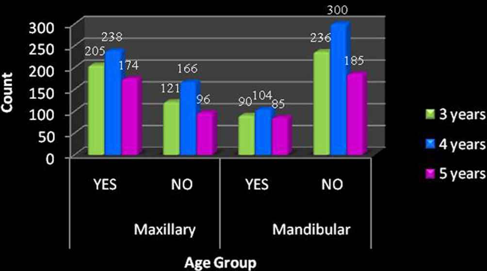
Age-wise distribution of primate spaces in maxillary and mandibular arch in children.
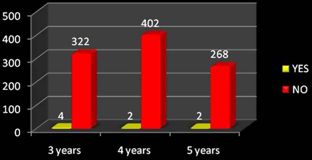
Prevalence of posterior cross-bite in children.
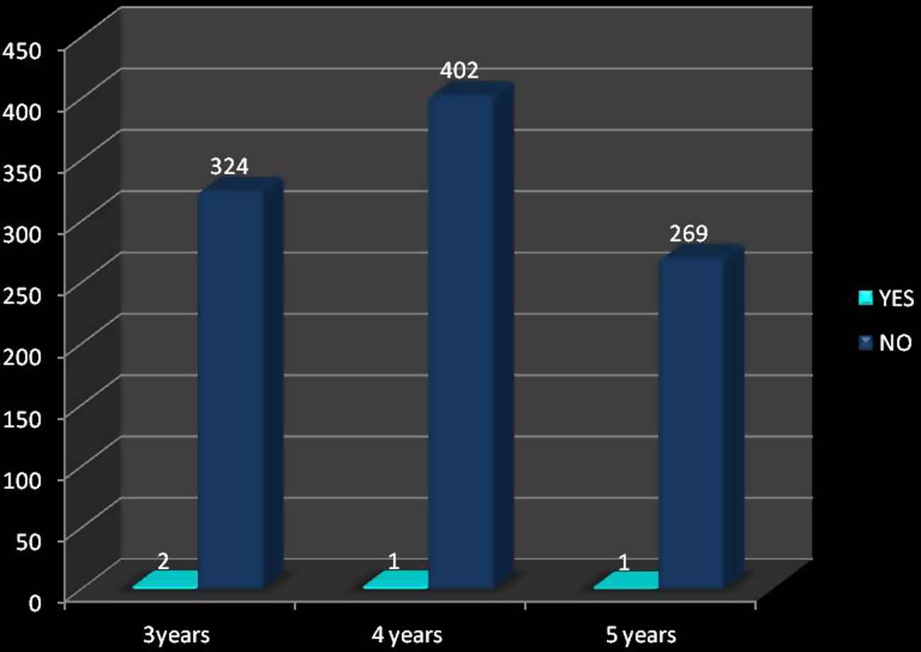
Prevalence of hypodontia in children.
Prevalence of fusion and gemination in children.
The early detection of the conditions that predispose one to develop a malocclusion in permanent dentition is very important. A thorough understanding of anterior and posterior changes occurring in occlusion between primary and early permanent dentition is crucial for a Pedodontist for early interceptive treatment.
3.1. Physiological and primate spaces
Physiological and primate spaces are developmental spaces in primary dentition; they play a very important role in the alignment of erupting permanent teeth and the establishment of occlusion. Absence of these spaces may be an expression of disproportion between the jaw and tooth size. In the present study, the prevalence of physiological spaces in maxillary and mandibular arches was 50.9% and 46.7%, respectively (Fig. 1). Also, primate spaces were present in 61.7% of children in the maxillary arch and 27.9% in the mandibular arch (Fig. 2). The study done by Bhayya and Shayagali goes well with the present study in which physiological spacing was the most prevalent characteristic in primary dentition with the prevalence of 35.4% in the maxillary and 25.7% in the mandibular arch. In the same study, a higher frequency of primate spaces was observed in the maxillary arch (47.6%) as compared with the mandibular arch (12.9%) [8]. The overall prevalence of physiological spaces and primate spaces in the present study was more in the maxilla than in the mandible, and the findings were in agreement with other studies as well [9,10].
3.2. Prevalence of anterior cross-bite
Anterior cross-bite is a malocclusion that may be diagnosed in growing patients, occurring in approximately 4–5% of the population in the primary or transitional dentition. In the present study, the prevalence of an anterior cross-bite was 0.1% (N = 1; age group 4 years), which is less in comparison with 5.5% in Nigerian children, 1.9% in Bagalkot children and 1.7% in Saudi children [8,11–13]. In such cases an early interceptive treatment is necessary to enhance the favorable growth and development of not only the occlusion, but also of the entire craniofacial complex [14,15].
3.3. Prevalence of posterior cross-bite
In the present study, unilateral posterior cross-bites were seen in 0.8% of the children (Fig. 3) which is less in comparison with 8.7% in Saudi Arabian children, 7% in Jordanian children, 7.1% in white children and 4.8% in Nigerian children [13,16–18]. Infante reported that the prevalence of posterior cross-bite was significantly greater in white children than in black and Indian children [1]. The difference in the prevalence rate of posterior cross-bite between the different cultures or ethnic groups may be due to the difference in the prevalence of habits which further influence the development of occlusion [11,19].
3.4. Prevalence of infra-occlusion
Infra-occlusion (submerged teeth) in relation to primary molar was not observed in the present study, although this variation has been reported in other populations with the frequency of 0.6% in Nigerians and 0.5% in Australian aborigines [17,20,21].
3.5. Prevalence of scissors bite
A buccal cross-bite with a whole segment of the upper teeth outside the lower arch is particularly termed as scissors-bite. In the present study, scissors-bite was not observed in the examined children similar to a study in Saudi children [17].
3.6. Hypodontia
Hypodontia refers to the congenital absence of one or multiple teeth. It is a rare anomaly in primary dentition. Hypodontia can be due to injury to the developing tooth germ, physical obstruction, disruption of the dental lamina, space limitations, or functional abnormalities of the dental epithelium or mesenchyme. Children who have hypodontia in the primary dentition have higher chances of having hypodontia in permanent dentition. It is rare for the primary teeth to be missing in both the maxillary and mandibular arches [22]. In the present study, hypodontia was seen in four children with the prevalence of 0.4% (Fig. 4) which is similar to that reported by Grahnen and Granath, but less than the 2% as reported by Chen [18,23]. Three of the four children with hypodontia had a missing mandibular central incisor with one of them showing the condition bilaterally. Also, hypodontia in relation to maxillary primary lateral incisor was found in a 3-year-old. Similarly, Ravn et al. reported that primary maxillary lateral incisors and mandibular incisors are the most frequently missing teeth [24]. In this study the clinically missing teeth were relied upon for hypodontia. The major drawback of this study was the lack of radiographic evaluation to confirm the congenitally missing teeth. It could be either un-erupted or impacted which can be ruled out only by a radiograph. The radiographic evaluation is necessary to confirm, hence, the children were asked to visit the Department of Pedodontics for a radiographic examination. Lack of radiographic evaluation in this study could have given an over-estimation of Hypodontia as many children could have pseudo-anodontia (in which teeth could not have erupted) and an under-estimation of hyperdontia (due to un-erupted supernumerary teeth in the jaw bones).
3.7. Fusion and gemination
Fusion is the union of two normally separated tooth germs, whereas gemination is an attempt at division of a single tooth germ. Dental fusion results in one tooth less than normal if the affected tooth is counted as 1, while gemination results in the normal number of teeth if the affected tooth is also counted as one. Gemination may occur in a tooth germ adjacent to a congenitally absent tooth, and this would be indistinguishable, clinically, from fusion. So the term “double teeth” or “double formation” is useful and includes both fusion and gemination. Fused teeth can be of two types: complete and incomplete. It is said to be complete when fusion begins before calcification due to which the crown incorporates the features of both the teeth involving their enamel, dentin, cementum and pulp, whereas it is incomplete when fusion occurs after calcification in which case a tooth can be found exhibiting separate crowns and fusion may be limited to the roots alone with pulp canals fused or separate. They may also contribute to esthetic concerns, space problems, occlusal disturbances and delayed eruption of permanent successors. It has been reported that approximately 50% of fused or geminated primary teeth result in abnormal permanent teeth like hypodontia, hence careful monitoring of the condition is recommended [23,25].
In the present study fusion was seen in three children and gemination was seen in two children. The combined prevalence of fusion and gemination (double teeth) in the present study is 0.5% (Fig. 5), which is comparable to other studies carried out in different ethnic groups (0.1–3%) [12,22,26,27].
Fusion in the primary teeth more frequently occurs unilaterally in the mandibular lateral incisor and canine region, whereas gemination mainly affects the primary maxillary lateral incisor. Similar findings were observed in the present study in which fusion was most prevalent in the mandible, whereas gemination was seen in the maxillary arch involving the primary right lateral incisor in a child in the 4-year-old age group. Fused teeth appear to occur more commonly than geminated teeth as seen in this study [25]. One rare finding was observed in the present study in which fusion between the primary right mandibular central and the lateral incisors and gemination of the primary right maxillary central incisor was seen in the same child.
4. Conclusion
- ▪
The prevalence of physiological spaces in the maxillary and mandibular arches was 50.9% and 46.7% respectively, whereas primate spaces were found in 61.7% of children in the maxillary arch and 27.9% in the mandibular arch.
- ▪
The prevalence of unilateral anterior and posterior cross-bite was 0.1% and 0.8%, respectively.
- ▪
The prevalence of hypodontia in the primary dentition was 0.4%.
- ▪
The prevalence of fusion and germination (double teeth) in the present study was 0.5%.
- ▪
In the primary dentition, double teeth (fusion and gemination) and hypodontia were the most common dental anomalies.
Conflict of interest
None declared.
References
Cite this article
TY  - JOUR
AU  - Seema Lochib
AU  - K.R. Indushekar
AU  - Bhavna Gupta Saraf
AU  - Neha Sheoran
AU  - Divesh Sardana
PY  - 2014
DA  - 2014/08/07
TI  - Occlusal characteristics and prevalence of associated dental anomalies in the primary dentition
JO  - Journal of Epidemiology and Global Health
SP  - 151
EP  - 157
VL  - 5
IS  - 2
SN  - 2210-6014
UR  - https://doi.org/10.1016/j.jegh.2014.07.001
DO  - 10.1016/j.jegh.2014.07.001
ID  - Lochib2014
ER  -